Lecture
A person’s motor activity has a very wide range - from muscle coordination required for rough manual work or moving the whole body in space, to subtle finger movements during operations that are performed under a microscope. Providing all types of motor activity is based on the movement of two streams of information. One stream originates at the periphery: in the sensing elements (receptors), which are located in the muscles, articular bags, tendon organs. Through the posterior horns of the spinal cord, these signals go up through the spinal cord and then into different parts of the brain .
Taken together, the signals from these structures form a special kind of sensitivity — proprioception. Although this information is not reflected in the mind of a person, thanks to her, the brain at every current time has a complete idea of the state of all its many muscles and joints. This information forms the pattern, or image, of the body. Without such an integrated education, a person could not plan and implement any movement. The body scheme is the initial basis for the implementation of any motor program. Its planning, construction and execution is related to the activity of the motor system.
In the motor system, the main information flow is directed from the motor zone of the cerebral cortex, the main center of arbitrary motion control, to the periphery, i.e. to the muscles and other organs of the musculoskeletal system, which carry out the movement.
The structures responsible for the nervous regulation of body position in space and movements are located in different parts of the central nervous system - from the spinal cord to the cerebral cortex. In their location, there is a clear hierarchy, reflecting the gradual improvement of motor functions in the process of evolution.
There are two main types of motor functions: maintaining the position (posture) and the movement itself. In everyday motor activity, it is quite difficult to separate them. Movement without simultaneously holding a pose is just as impossible as holding a pose without movement. (see fig.)
The structures responsible for the nervous regulation of posture and movements are located in different parts of the central nervous system - from the spinal cord to the cerebral cortex. In their location, there is a clear hierarchy, reflecting the gradual improvement of motor functions in the process of evolution.
Below is a general plan for the organization of the motor system (see Table 10.1).
Table 10.1
| Structure | Function performed by an isolated structure | The role of structure in the implementation of the movement |
| Subcortical and cortical motivational zones | Motivation for action | Plan |
| Associative zones of the cortex | Action plan | Plan |
| Basal ganglia cerebellum |
Schemes of targeted movements | Program |
| Thalamus Motor cortex |
(Acquired and congenital) | Program and its implementation |
| Brain stem | Posture adjustment | Performance |
| Spinal neurons | Mono- and polysynaptic reflexes | Performance |
| Motor units | Muscle length and tension | Performance |
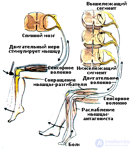
The lowest level in the organization of movement is associated with the motor systems of the spinal cord. In the spinal cord between sensitive neurons and motor neurons, which directly control the muscles, intercalated neurons are located, which form many contacts with other nerve cells. From the excitation of intercalary neurons depends on whether this or that movement is facilitated or inhibited. The neural circuits, or reflex arcs, underlying the spinal reflexes, are anatomical structures that provide the simplest motor functions. However, their activity largely depends on the regulatory influences of the centers located above.
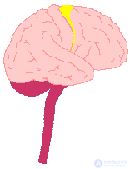 Higher motor centers are located in the brain and provide for the construction and regulation of movements. Motor acts aimed at maintaining posture, and their coordination with targeted movements is carried out mainly by the brain stem structures, while at the same time targeted movements themselves require the participation of higher nervous centers. The urge to action associated with the excitation of subcortical motivational centers and associative zones of the cortex , forms a program of action. The formation of this program is carried out with the participation of the basal ganglia and the cerebellum acting on the motor cortex through the nuclei of the thalamus (see Video). Moreover, the cerebellum plays a pivotal role in the regulation of posture and movements, and the basal ganglia constitute the link between the associative and motor regions of the cerebral cortex.
The motor, or motor, cortex is located directly anterior to the central sulcus. In this zone, the muscles of the body are represented topographically, i.e. each muscle has its own part of the area. Moreover, the muscles of the left half of the body are represented in the right hemisphere, and vice versa.
Motor pathways leading from the brain to the spinal cord are divided into two systems: pyramidal and extrapyramidal . Starting in the motor and sensory zones of the cerebral cortex, most of the fibers of the pyramidal tract are directed directly to the efferent neurons in the anterior horns of the spinal cord. The extrapyramidal tract, which also goes to the anterior horns of the spinal cord, transmits to them efferent impulses processed in a complex of subcortical structures (basal ganglia, thalamus, cerebellum).
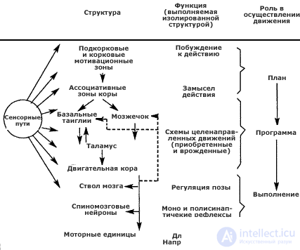 |
The general plan of the organization of the motor system (according to J. Dudella et al., 1985). The most important motor structures and their main relationships are shown in the left column. For simplicity, all sensitive paths are combined together (circle on the left). The middle column lists the most important and well-established functions found when studying each of these structures separately. The right column indicates how these functions are related to the occurrence and execution of motion. It should be noted that the basal ganglia and the cerebellum are located at the same level, and the motor cortex is involved in the transformation of the movement program in its implementation |
All variety of forms of movement of animals and humans is based on the physical laws of the movement of bodies in space. When classifying movements, it is necessary to take into account the specific objective functions that the propulsion system must perform.
The hierarchy of the levels of brain control movements also depends on the requirements for the structure of the movement. It is established that the subcortical level is associated with a set of innate or automated programs.
 Automated and arbitrary movements. The problem of separation of these categories of movement is complex. In many cases, the line between an automated and arbitrarily controlled action is very mobile. Moreover, the essence of learning motor skills is the transition from a constantly controlled chain of more or less consciously shared motor actions to an automated, continuous “kinetic melody”, which is performed with much lower energy costs. At the same time, a small change in at least one of the components of the automated skill is enough to stop this skill from being fully automated, and arbitrary regulation intervention is required.
In order to avoid the difficulties arising from attempts to divide motor acts into "automatic" and "volitional", the English neuropathologist H. Jackson at the beginning of the century proposed a hierarchical classification of all motor acts (ie, movements and their complexes) from the "fully automatic" to "completely arbitrary." This classification proves useful now. For example, breathing is a largely automatic set of movements of the chest and muscles of the shoulder girdle, which persists even during the deepest sleep and in the state of anesthesia, when all other movements are completely suppressed. If with the help of the same muscles a cough reflex or torso movement is performed, then a similar motor act is “less automatic”, and when singing or speaking these muscles are already involved in “completely non-automatic” movement. It is also clear from this example that “more automatic” movements are mainly associated with innate central behavioral programs, while “less automatic” or “completely arbitrary” movements appear in the process of accumulating life experience.
Orientational movement. The system of movements of this type is associated with the orientation of the body in space and with the installation of the senses in a position that provides the best perception of an external stimulus. An example of the first is the function of maintaining balance, the second is the movement of fixing the gaze. Fixing the gaze is performed mainly by the oculomotor system. The image of a fixed or moving object is fixed in the most sensitive field of the retina. Coordination of eye and head movement is regulated by a special system of reflexes.
Pose control. The pose of the body is determined by the totality of the angles formed by the joints of the human body as a result of orientation in the field of aggression. The mechanism of posture consists of two components: the fixation of certain body positions and limbs and the orientation of body parts relative to the external coordinates (maintaining balance). The original body posture imposes some restrictions on subsequent movement. The lower mechanisms of posture control include spinal , cervical adjusting and some other reflexes, and the higher mechanisms are mechanisms of forming a "body pattern".
The term "body diagram" refers to the system of generalized sensitivity of one's own body at rest and during movement, spatial coordinates and interrelations of individual parts of the body. A common "map" of the body for each hemisphere of the brain is usually presented as a "homunculus". The sensitivity of the whole body, which is topographically distributed over the surface of the cortex, forms the basis from which integral functional blocks of large parts of the body are formed. These integrative processes are completed in an adult organism and are an encoded description of the relative position of body parts, which are used when performing automated stereotyped movements.
The base of these processes is the anatomically fixed “map” of the body, therefore such processes are only the basis of the static body image. To form it, it is necessary to correlate this information with the position of the body relative to the force of gravity and the relative position of the functional blocks of the body in the system of three spatial planes. The vestibular system perceives the movement of the entire body back and forth, left and right, up and down, and the corresponding information enters the parietal areas of the cortex, where it is combined with information from the musculoskeletal system and skin. It also receives impulses from the internal organs, which also participate in the creation at the unconscious level of a special psychophysiological formation - a static body image.
Thus, the static body image is a system of intracerebral connections, based on innate mechanisms and improved and refined in ontogenesis. While performing this or that activity, a person changes the interposition of body parts, and while learning new motor skills, he forms new spatial models of the body, which form the basis of a dynamic body image. In contrast to the static, the dynamic image of the body matters only for this particular point in time and a certain situation, when it changes, it is replaced by a new one. The dynamic image is based on current impulses from sensitive elements of the skin, muscles, joints and the vestibular apparatus. It is possible that the speed and accuracy of the formation of a dynamic body image is a factor that determines a person’s ability to quickly master new motor skills.
In the brain, there is a constant interaction of this and another body image, a comparison of the dynamic image with its static analogue is carried out. As a result, a subjective feeling of posture is formed, reflecting not only the position of the body at a given time, but also its possible changes in the immediate future. If coordination is not achieved, then active posture adjustment mechanisms come into play. So, in order to change the pose, it is necessary to compare the static image of the body encoded in memory with its specific variation - the dynamic image of the body.
 Control of locomotion. The term locomotion means moving a body in space from one position to another, for which a certain expenditure of energy is necessary. The efforts developed at the same time must overcome, first of all, the force of gravity, the resistance of the environment and the forces of inertia of the body itself. Locomotion is influenced by the nature and terrain. During locomotion, the body must constantly maintain balance.
Typical examples of locomotion are walking or running, which are distinguished by stereotypical movements of the limbs, and for each form of locomotion there are two phases of a step: the support phase and the transfer phase. A person’s walking is characterized by gait, i.e. the inherent features of movement on the surface. Gait is estimated by the method of time distribution of cyclic movements of the limbs, the duration of the supporting phase and the sequence of movement of the supporting limbs.
A chain of neurons has been found in the spinal cord that serves as a walking generator. She is responsible for the alternation of the periods of excitation and inhibition of various motoneurons and can work in automatic mode. The elementary unit of such a central generator is a generator for one limb. It is possible that each muscle that controls one joint has its own generator. When a person moves, such generators work in a single mode, exerting a stimulating effect on each other.
As you know, the spinal cord is under the continuous control of higher motor centers.
A very important role in this control is played by the cerebellum , which provides correction and accuracy of limb setting based on a comparison of information about the work of the spinal generator and the actual parameters of the movements. It is assumed that the cerebellum programs each next step on the basis of information about the previous one. Another important level of the brain, where information about the nature of the movement is directed, is the cerebral hemispheres with their thalamic nuclei, the striopallidar system and the corresponding areas of the cerebral cortex.
Feedback. Feedback is of great importance at these levels of locomotion control, i.e. information about the results of the movement being performed. It comes from the motor apparatus to the corresponding brain centers. Many movements are constantly corrected, thanks to the indications of the corresponding sensory sensors located in the skeletal muscles and transmitting information to different parts of the brain right down to the cortex. Movements based on innate coordinates require less feedback from the locomotor apparatus. Along with this, all new forms of movement, which are based on the formation of new coordination relations, are entirely dependent on feedback from the locomotor system.
It is very important that sensory corrections can change the nature of movement in the course of its implementation. Without this mechanism, a person would not have had the opportunity to master new locomotor acts (and not only “locomotor masterpieces”, which are demonstrated by masters of artistic gymnastics, but also by simpler ones, such as cycling). The essence of the matter is that sensory corrections serve to clarify the dynamic image of the body, bringing it as close as possible to the requirements of movement.
So, simple movements (for example, spasmodic eye movements or fast movements of the extremities) are performed with little or no proprioceptive feedback in a rigid “sealed” program. Any complex movement requires preliminary programming. For complex movements, it is very important to compare reverse afferentation with the sensory image of movement that forms in the program. These influences are transmitted to programming devices via internal feedback channels, which includes all the processes of reorganization of the motor program depending on intracentral influences.
Следует особо подчеркнуть, что с помощью обратной связи кора информируется не об отдельных параметрах движений, а о степени соответствия предварительно созданной двигательной программы тому наличному движению, которое достигается в каждый момент времени. Одним из важнейших каналов такой внутренней обратной связи и выступают медиальные лемниски.
A distinctive feature of manipulator movements is their dependence on the central program, therefore the frontal cortex, the basal ganglia and the cerebellum play a leading role in the implementation. The leading role in the programming of fast manipulatory movements belongs to the cerebellar system, and in the programming of the slow, to the basal ganglia.
The hierarchy of forms of motor activity (according to N. A. Bernstein). The most complete problem of the hierarchical organization of human movements in the context of active adaptive behavior was posed and developed in the works of the outstanding Russian physiologist N.А. Bernstein. He developed a theory of motion building levels. Moreover, by levels he understood the morphological parts of the nervous system: the spinal cord and the medulla, the subcortical centers and the cortex of the cerebral hemispheres. Each level has its own type of movements. Total N.A. Bernstein singled out five levels: A, B, C, D, E.
1. Level A is the most evolutionarily ancient and maturing spinal spinal level. In humans, it has no independent value, but it determines muscle tone. и участвует в обеспечении любых движений совместно с другими уровнями. Есть некоторые формы двигательной активности, которые осуществляются только за счет данного уровня (к их числу относятся непроизвольные примитивные движения, например, дрожание пальцев, стук зубов от холода). Этот уровень начинает функционировать с первых недель жизни новорожденного.
2. Уровень В- thalamopalidar level, provides processing of signals from the muscular-articular receptors, which report the relative positioning of body parts. This level takes part in the organization of movements of a more complex type, which, however, do not require taking into account the peculiarities of the external space. It can be arbitrary movements of the face and body - facial expressions and pantomime, freestyle gymnastics, etc. This level begins to function in the second half of the child’s life.
3. Level C- defined as the level of the spatial field or pyramidal-streaky level. This level receives information about the state of the environment from exteroreceptor analyzers. Therefore, this level is responsible for the construction of movements adapted to the spatial properties of objects - to their shape, position, weight, and other features. Among them are all types of locomotion (movement), fine hand motor skills and others. This is the level in which the cortex takes part along with the subcortical structures. Therefore, its maturation, starting very early - in the first year of life, continues throughout childhood and even adolescence.
4. Level D- the level of substantive action. It operates with the obligatory participation of the cortex (parietal and premotor zones) and ensures the organization of actions with objects. This is a specifically human level of organization of motor activity, since it includes all types of tool actions and manipulative movements. A characteristic feature of the movements of this level is that they not only take into account spatial features, but also are consistent with the logic of the use of the object. These are not only movements, but also to a much greater degree of action, because the motor programs used here consist of flexible interchangeable links. Since this level is ensured by the coordinated activity of different zones of the cortex, its functionality will be determined by the dynamics of maturation as zones themselves,and age features of interzone interaction.
5. Уровень Е — высший уровень организации движений, обеспечивает интеллектуализированные двигательные акты: работу артикуляционного аппарата в звучащей речи, движения руки при письме, а также движения символической или кодированной речи (язык жестов глухонемых, азбука Морзе). Нейрофизиологические механизмы этого уровня обеспечиваются высшими интегративными возможностями коры больших полушарий, поэтому созревание коры, как и в предыдущем случае, имеет решающее значение для его функционирования.
Программирование движений. Каждому целенаправленному движению предшествует формирование программы, которая позволяет прогнозировать изменения внешней среды и придать будущему движению адаптивный характер. Результат сличения двигательной программы с информацией о движении, передающейся по системе обратной связи, является основным фактором перестройки программы. Последнее зависит от мотивированности движения, его временных параметров, сложности и автоматизированности (см. Видео).
Motivation determines the overall strategy of the movement. Each specific motor act is often a step towards the satisfaction of a particular need. Biological motivations lead to the launch of either hard, largely genetically determined motor programs, or form new complex programs. However, motivation determines not only the purpose of the movement and its program, it also determines the dependence of movement on external stimuli. The feedback here is the satisfaction of need.
The motor command determines how the programmed movement will be performed, i.e. what is the distribution in time of those efferent volleys sent to the motoneuronsspinal cord, which will cause the activation of various muscle groups. In contrast to programs, teams of movement must exactly correspond to the functional state of the skeletal-motor system itself as a direct executor of these commands. Direct movement control is determined by the activity of the motor zone of the cortex, striatum and cerebellum. The striatum is involved in the transformation of the “intention to act” into the appropriate “command signals” to initiate and control movements.
Особую роль в программировании движения играют ассоциативные системы мозга, и в первую очередь таламопариетальная ассоциативная система. Во-первых, именно она участвует в формировании интегральной схемы тела. При этом все части тела соотносятся не только друг с другом, но и с вестибулярными и зрительными сигналами. Во-вторых, она регулирует направление внимания к стимулам, поступающим из окружающей среды так, чтобы учитывалась ориентация всего тела относительно этих стимулов. Эта система "привязана" к настоящему моменту времени и к анализу пространственных взаимоотношений разномодальных признаков.
Таламофронтальная ассоциативная система отвечает за переработку информации о мотивационом состоянии и происходящих в организме вегетативных изменениях. Фронтальная ассоциативная область коры опосредует мотивационные влияния на организацию поведения в целом благодаря связям с другими ассоциативными областями и подкорковыми структурами. Таким образом, фронтальные отделы коры больших полушарий, контролируя состояние внутренней среды организма, сенсорные и моторные механизмы мозга, обеспечивают гибкую адаптацию организма к меняющимся условиям среды (см. Хрестомат. 10.1).
Функциональная структура произвольного движения. Из вышеизложенного следует, что в обеспечении любого движения принимают участие разные компоненты, поэтому один из главных вопросов состоит в том, каким образом обеспечивается единовременность команды, поступающей к исполнительным аппаратам. Независимо от стратегии и тактики конкретного движения, основная задача системы, обеспечивающей программу, заключается в координации всех компонентов команды.
ЦНС располагает некоторым числом генетически закрепленных программ (например, локомоторная программа шагания, базирующаяся на активности спинального генератора). Такие простые программы объединяются в более сложные системы типа поддержания вертикальной позы. Подобное объединение происходит в результате обучения, которое обеспечивается благодаря участию передних отделов коры больших полушарий.
The most complex and phylogenetic of the youngest is the ability to form a sequence of movements and to anticipate its implementation. The solution to this problem is connected with the frontal associative system, which stores and stores such sequences of movements. The highest reflection of this coding in humans is verbalization, or verbal accompaniment, of the basic concepts of movement.
The universal pattern of the motion control system is the use of feedback. This includes not only proprioceptive feedback from the onset of movement, but also activation систем поощрения или наказания. Кроме того, включается и внутренняя обратная связь, т.е. информация об активности нижележащих уровней двигательной системы, или эфферентная копия самой двигательной команды. Этот вид обратной связи необходим для выработки новых двигательных координаций. Для движений различной сложности и скорости обратная связь может замыкаться на разных уровнях. Поэтому оба типа управления — программирование и слежение — могут сосуществовать в системе управления одним и тем же движением.
В заключение целесообразно привести высказывание выдающегося физиолога Н.А. Бернштейна о том, что движения "...ведет не пространственный, а смысловой образ и двигательные компоненты цепей уровня действий диктуются и подбираются по смысловой сущности предмета и того, что должно быть проделано с ним".
Электрофизиологические методы используются для изучения разных сторон двигательной активности, и в первую очередь тех из них, которые недоступны прямому наблюдению. Ценную информацию о физиологических механизмах организации движения дают методы оценки взаимодействия зон коры мозга, анализ локальной ЭЭГ и потенциалов, связанных с движением, а также регистрация активности нейронов.
Исследование межзональных связей биопотенциалов мозга позволяет проследить динамику взаимодействия отдельных зон коры на разных этапах выполнения движения, при обучении новым двигательным навыкам, выявить специфику межзонального взаимодействия при разных типах движений.
Spatial synchronization (PS) , i.e. The synchronous dynamics of electrical oscillations recorded from different points of the cerebral cortex reflects a state of the brain structures that facilitates the spread of arousal and creates conditions for inter-zone interaction. The method of registration of PS was developed by an outstanding domestic physiologist M.N. Livanov.
Studies of the rhythmic components of the EEG of individual zones and their spatial-temporal relationships in humans during the execution of voluntary movements gave a real opportunity to approach the analysis of the central mechanisms of functional interactions, which are formed at the system level during motor activity. The correlation analysis of the EEG recorded during the execution of rhythmic movements showed that in a person in the cortical organization of the movements, not only the centers of the motor cortex, but also the frontal and lower dark areas are involved.
Training in voluntary movements and their training cause the redistribution of intercentral correlations of cortical biopotentials. At the beginning of training, the total number of centers involved in joint activities increases dramatically, and the synchronous relations of the rhythmic components of the EEG motor zones with the front and rear associative regions are strengthened. As the movement is mastered, the overall level of the PS is significantly reduced, and, on the contrary, the links between the motor zones and the lower ones are strengthened. 
It is important to note that in the process of learning, the rhythmic composition of the biopotentials of different zones of the cortex occurs: slow rhythms that coincide in frequency with the rhythm of the movements are started to be recorded in the EEG. These rhythms in the human EEG are called "labeled". The same marked oscillations were found in preschool children when they made rhythmic movements on an ergograph.
Systematic studies of human EEG during cyclic (periodically repeated) and acyclic motor activity revealed significant changes in the dynamics of the electrical activity of the cerebral cortex. In the EEG, both local and distant synchronization of biopotentials are amplified, which results in an increase in the power of periodic components, changes in the frequency spectrum of auto and cross correlograms, in a certain alignment of the maxima of the frequency spectra and coherence functions at the same frequency.
PS and reaction time. Reaction time - one of the most simple motor indicators. Therefore, of particular interest is the fact that even a simple motor reaction can have different physiological correlates depending on the increase or reduction of its duration. So, when comparing the picture of intercentral correlation relations of the spectral components of the brain EEG with the time of a simple motor reaction, it turned out that the restructuring of the space-time relations of the EEG associative zones is associated with the response time to a given stimulus. During fast reactions in a healthy person, most often high correlations of the biopotentials occurred in both lower tempered regions (somewhat more with the left hemisphere of the brain). If the reaction time increased, this was accompanied by synchronization of the biopotentials in the frontal regions of the cortex and the lower-tempered area of the left hemisphere was excluded from the interaction. In addition, a relationship was found between the values of the phase shifts of the alpha rhythm recorded in the frontal, precentral, and occipital regions of the brain and the speed of a simple motor reaction.
It is important to note that the enhancement of synchronization of biopotentials occurs in a person already in the pre-working state in the process of concentration before the motor action, as well as in the mental performance of movements.
PS and the specifics of the movement. In addition to the non-specific enhancement of the PS biopotentials, its pronounced selective increase between the cortical zones directly involved in the organization of a specific motor act was noted. For example, the greatest similarity in electrical activity is established: in the movement of the arms, between the frontal region and the motor representation of the muscles of the upper limbs; when the legs move, it is between the frontal area and the motor representation of the muscles of the lower limbs. With precision actions that require fine spatial orientation and visual control (shooting, fencing, basketball), the interactions between the visual and motor areas are enhanced.
The complex dynamics of the PS biopotentials of various brain areas in athletes during various exercises was revealed and the dependence of the increase in the interaction of rhythmic components of the EEG on the mode of motor activity, on the skills of athletes, on the person’s ability to solve tactical tasks, on the complexity of the situation was shown. Thus, in highly qualified athletes, intercenter interactions are expressed much more intensely and localized more clearly. It also turned out that more complex motor tasks require for their successful solution a higher level of spatial synchronization of EEG rhythms, and the time required for solving tactical tasks correlates with the rate of increase of intercentral interactions. In this case, the motor response follows after the maximum synchronization of the biopotentials in the cerebral cortex is reached.
Taken together, studies of the brain's PS biopotentials in humans made it possible to establish that, when performing simple and complex motor acts, different centers of the brain enter into interactions, forming complex systems of interconnected zones with foci of activity not only in projection but also in associative areas, especially the frontal and low-grade These intercenter interactions are dynamic and change in time and space as the motor act proceeds.

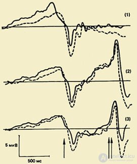 One of the important directions in the study of the psychophysiology of the motor act is the study of the complex of oscillations of the potentials of the brain associated with movements (PMSD). The significance of this phenomenon for understanding the physiological mechanisms of movement organization is very great, because studying IMSD allows us to reveal a hidden sequence of processes occurring in the cerebral cortex during the preparation and execution of movement, and to time these processes, i.e. set the time limits for their flow.
The composition of the PMSD. For the first time, this complex, reflecting the processes of preparing, executing and evaluating the movement, was registered in the 1960s. It turned out that the movement is preceded by a slow negative oscillation - the potential of readiness (PG). It begins to develop in 1.5 - 0.5 seconds before the start of the movement. This component is recorded mainly in the central and frontal-central leads of both hemispheres. 500-300 ms before the start of motion, the PG becomes asymmetrical - its maximum amplitude is observed in the precentral area, which is contralateral to motion. In about half of the adult test subjects, a small amplitude positive component is recorded against the background of this slow negative oscillation, shortly before the start of the movement. He received the name "premotor positivity" (PMP). The next negative oscillation, rapidly increasing in amplitude, the so-called motor potential (MP), begins to develop 150 ms before the start of the movement and reaches a maximum amplitude above the motor representation of the moving limb in the cerebral cortex. This complex of potentials ends with a positive component approximately 200 ms after the start of movement.
The functional significance of the components. It is believed that the readiness potential (GHG) arises in the motor cortex and is associated with the planning and preparation processes of the movement. It belongs to the class of slow negative fluctuations of the brain potential, the occurrence of which is explained by the activation of the neuronal elements of the corresponding areas of the cortex.
Hypotheses regarding the functional value of the PMP are different.
This oscillation is considered both as a reflection of the supply of the central team from the cortex to the muscles, and as a result of relaxation of the cortex after the completion of a certain stage of movement organization, and as a reflection of the processes of suppression of the associated movements of the other limb, and as feedback from muscle afferents. Currently, some authors believe that PMP is only a reflection of the beginning of the motor potential.
When registering MPs in monkeys as part of MPs, two subcomponents were identified. The first subcomponent is correlated with the activation of the motor cortex associated with the initiation of movement (synaptic activity of pyramidal neurons), and the second with the activation of 2.3 and 4 fields according to Brodmann . The registration of MP in a person with epilepsy allowed us to distinguish three components in it. The first component was called initiation potential. It has a high amplitude and occurs after the start of movement in the precentral contralateral cortex. The second one, which appears after the start of the myogram, is more localized in the contralateral somatosensory field and can be associated with both the initiation of movement and sensory feedback. The third component reflects the impulses coming from the muscle afferents into the cortex.
The positive potential following the MP is considered as a reflection of inverse afferentation coming from peripheral receptors, upward activity from motor centers, a comparison operation between a motor program and a neuronal picture of its performance or relaxation processes of the cortex after the movement has been completed.
Waiting wave In addition to PMSD, another electrophysiological phenomenon is described, which is inherently close to the potential of readiness. This is a negative potential fluctuation recorded in the anterior cerebral cortex in the period between the action of the warning and the trigger (requiring a reaction) signals. This oscillation has a number of names: waiting wave, E-wave, conditional negative deviation (UNF). The E-wave occurs 500 ms after the warning signal, its duration increases with increasing interval between the first and second stimuli. The amplitude of the E-wave increases in direct proportion to the speed of the motor reaction to the starting stimulus. It increases with tension of attention and increase of volitional effort, which indicates the connection of this electrophysiological phenomenon with the mechanisms of arbitrary regulation of motor activity and behavior in general.
Functional cortical columns. In the motor zone of the cortex in humans there are so-called giant Betz pyramidal cells , which are organized in separate columns. Pyramidal cells that perform similar functions are located next to each other, otherwise it would be difficult to explain the exact somatotopic organization of the cortex. Such motor columns are able to excite or inhibit a group of functionally homogeneous motoneurons .
Registration of the activity of single pyramidal cells with the help of implanted microelectrodes in animals performing various movements, made it possible to establish a fundamentally important fact. The neurons of the cortex that regulate the activity of any muscle are not concentrated within just one column. The motor column is largely a functional association of neurons that regulate the activity of several muscles acting on a particular joint. Thus, in the columns of the pyramidal neurons of the motor cortex, not so much muscle as movement is represented.
Neural codes of motor programs. The coding of information in a neuron is carried out by the frequency of its discharges. Analysis of the impulse activity of neurons in the development of various motor programs in animals showed that neurons from different parts of the motor system participate in their construction, while performing specific functions. According to some ideas, the inclusion of motor programs occurs due to the activation of so-called command neurons. Command neurons are, in turn, under the control of higher cortical centers. The inhibition of the command neuron leads to the cessation of the program it controls, but excitement, on the contrary, to the activation of the nervous circuit and the actualization of the motor program.
The involvement of command neurons in the whole brain activity is determined by the current motivation and a specific motor program aimed at satisfying this motivation. The motor program, in order to be adaptive in nature, must take into account all the signal-significant components of the external environment relative to which the targeted movement takes place, i.e. built on the principle of multisensory convergence .

Comments
To leave a comment
Psychophysiology
Terms: Psychophysiology