Lecture
In this section, systematics, registration methods and the importance of physiological indicators related to human mental activity will be presented. Psychophysiology is an experimental discipline, therefore the interpretational possibilities of psychophysiological research are largely determined by the perfection and diversity of the methods used. The correct choice of methodology, the adequate use of its indicators and the interpretation of the results obtained, which correspond to the resolving possibilities of the methodology, are the conditions necessary for conducting a successful psychophysiological study.
The central place among the methods of psychophysiological research is occupied by various ways of recording the electrical activity of the central nervous system, and first of all the brain.
 Electroencephalography is a method of recording and analyzing electroencephalogram (EEG), i.e. total bioelectric activity, abstracted from both scalp and deep brain structures . The latter is possible only in a clinical setting.
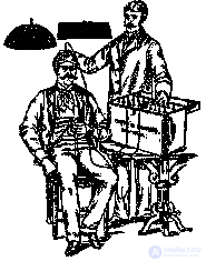
In 1929, the Austrian psychiatrist H. Berger discovered that brain waves can be recorded from the surface of the skull. He established that the electrical characteristics of these signals depend on the state of the subject. The most noticeable were synchronous waves of relatively large amplitude with a characteristic frequency of about 10 cycles per second. Berger called them alpha waves and contrasted them with high-frequency "beta waves" , which occur when a person becomes more active. The discovery of Berger led to the creation of an electroencephalographic method for studying the brain, consisting in recording, analyzing and interpreting the biological currents of the brain of animals and humans.
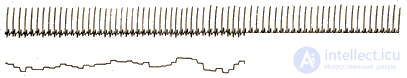
One of the most striking features of an EEG is its spontaneous, autonomous nature. Regular electrical activity of the brain can be fixed in the fetus (ie, before the birth of the body) and stops only with the onset of death. Even with deep coma and anesthesia, there is a special characteristic pattern of brain waves.
Today, EEG is the most promising, but still the least deciphered data source for the psychophysiologist.
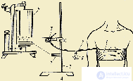
Registration conditions and methods of EEG analysis. The stationary complex for recording EEG and a number of other physiological indicators includes a soundproofed shielded camera, an equipped place for the test, mono-channel amplifiers, recording equipment (black-and-white encephalograph, multi-channel tape recorder). Usually, 8 to 16 EEG recording channels are used from different parts of the skull surface at the same time. EEG analysis is carried out both visually and with the help of computers. In the latter case, special software is required.
It should be emphasized that such a division into groups is more or less arbitrary; it does not fit any physiological categories. Slower frequencies of the electrical potentials of the brain have been registered up to periods of the order of several hours and days. Recording on these frequencies is performed using a computer.
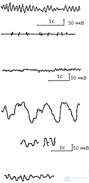 |
The main rhythms and parameters of the encephalogram. 1. Alpha-wave - a single two-phase oscillation of the potential difference with a duration of 75-125 ms., In form, approaches a sinusoidal one. 2. Alpha-rhythm - rhythmic fluctuation of potentials with a frequency of 8-13 Hz, expressed more often in the back of the brain with eyes closed in a state of relative rest, the average amplitude is 30-40 μV, usually modulated in the spindle. 3. Beta-wave - a single two-phase oscillation of potentials with a duration of less than 75 ms. and an amplitude of 10-15 μV (no more than 30). 4. Beta rhythm - rhythmic fluctuation of potentials with a frequency of 14-35 Hz. Better expressed in the fronto-central areas of the brain. 5. Delta-wave - a single two-phase oscillation of the potential difference with a duration of more than 250 ms. 6. Delta rhythm - rhythmic fluctuation of potentials with a frequency of 1-3 Hz and amplitude from 10 to 250 mV or more. 7. Theta wave - a single, often two-phase oscillation of the potential difference with a duration of 130-250 ms. 8. Theta rhythm is a rhythmic fluctuation of potentials with a frequency of 4-7 Hz, more often two-way synchronous, with an amplitude of 100-200 µV, sometimes with spindle-shaped modulation, especially in the frontal area of the brain. |
 Another important characteristic of the brain's electrical potentials is amplitude, i.e. oscillation magnitude. The amplitude and frequency of oscillations are related to each other. The amplitude of high-frequency beta waves from one and the same person can be almost 10 times lower than the amplitude of slower alpha waves.
The location of the electrodes is important when recording EEGs, and the electrical activity simultaneously recorded from different points of the head can vary greatly. When recording EEG using two main methods: bipolar and monopolar. In the first case, both electrodes are placed in electrically active points of the scalp, in the second one of the electrodes is located at a point that is conventionally considered electrically neutral (earlobe, nose bridge). In bipolar recording, an EEG is recorded, representing the result of the interaction of two electrically active points (for example, frontal and occipital leads), and in monopolar recording, the activity of any one lead is relatively electrically neutral (for example, frontal or occipital lead relative to the earlobe). The choice of one or another version of the record depends on the objectives of the study. In research practice, the monopolar registration option is more widely used, since it allows studying the isolated contribution of a particular brain area to the process under study.
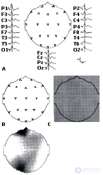
The International Federation of Electroencephalography Societies adopted the so-called "10-20" system, which allows to accurately indicate the location of the electrodes. In accordance with this system, the distance between the middle of the nose bridge (nasion) and the solid bone tubercle at the nape (inion), as well as between the left and right ear pits, is accurately measured for each person tested. Possible points of location of the electrodes are separated by intervals of 10% or 20% of these distances on the skull. At the same time, for the convenience of registration, the entire skull is divided into areas marked with letters: F - frontal, O - occipital region, P - parietal, T - temporal, C - region of the central sulcus. Odd numbers of places of assignment belong to the left, and even - to the right hemisphere. The letter Z - denotes the lead from the top of the skull. This place is called Vertex and is used especially frequently (see Chrestomat. 2.2).
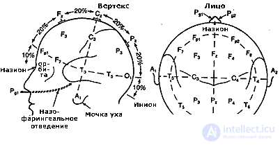 |
System 10-20 (Jasper, 1958). The location of the electrodes on the surface of the head: F - the frontal part; C - central; P - parietal; T - temporal; O - occipital. Odd indices - the left half of the head, even indices - the right, Z - the middle line |
Clinical and static methods of studying EEG. From the moment of their appearance, two approaches to EEG analysis have emerged and continue to exist as relatively independent: visual (clinical) and statistical.
Visual (clinical) EEG analysis is used, as a rule, for diagnostic purposes. The electrophysiologist, relying on certain methods of such an EEG analysis, solves the following questions: does the EEG comply with generally accepted standards of the norm; if not, what is the degree of deviation from the norm, whether the patient shows signs of focal brain damage and what is the localization of the lesion. Clinical analysis of EEG is always strictly individual and is primarily of a qualitative nature. Despite the fact that there are generally accepted in the clinic techniques for describing an EEG, the clinical interpretation of an EEG largely depends on the experience of the electrophysiologist, his ability to “read” the electroencephalogram, highlighting hidden and often very variable pathological signs in it.
It should, however, be emphasized that in wide clinical practice, gross macroscopic abnormalities or other distinct forms of EEG pathology are rare. Most often (70-80% of cases) there are diffuse changes in the bioelectrical activity of the brain with symptoms that are difficult to formally describe. Meanwhile, it is precisely these symptoms that may be of particular interest for the analysis of the contingent of subjects who belong to the group of so-called "minor" psychiatry - the states bordering on the "good" norm and apparent pathology. It is for this reason that special efforts are now being made to formalize and even develop computer programs for analyzing clinical EEG.
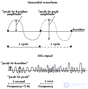
Statistical methods for the study of electroencephalograms assume that the background EEG is stationary and stable. In the overwhelming majority of cases, further processing relies on the Fourier transform, the meaning of which is that a wave of any complex shape is mathematically identical to the sum of sinusoidal waves of different amplitude and frequency.
The Fourier transform allows you to convert the background EEG wave pattern into a frequency one and establish the power distribution for each frequency component. With the help of the Fourier transform, the most complex in form of EEG oscillations can be reduced to a number of sinusoidal waves with different amplitudes and frequencies. On this basis, new indicators are highlighted, expanding the meaningful interpretation of the rhythmic organization of bioelectric processes.
For example, a special task is the analysis of the contribution, or relative power, of different frequencies, which depends on the amplitudes of the sinusoidal components. It is solved by building power spectra. The latter is a set of all values of the power of the rhythmic components of the EEG, calculated with a certain discretization step (in the amount of tenths of a hertz). The spectra can characterize the absolute power of each rhythmic component or relative, i.e. the severity of power of each component (in percent) relative to the total power of the EEG in the analyzed section of the record.
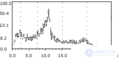 |
Individual EEG spectrum at rest (according to D. Lykken et al., 1974). The abscissa is the frequency in Hz., And the ordinate is the spectral density on a logarithmic scale. The figure clearly shows that the maximum value of the spectral power falls on the frequency of the alpha rhythm |
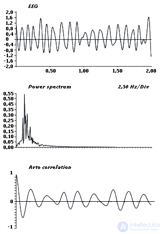 The EEG power spectra can be further processed, for example, by correlation analysis, and the auto- and cross-correlation functions, as well as coherence , which characterize the measure of synchrony of the EEG frequency ranges in two different leads , are calculated. Coherence ranges from +1 (completely matching waveforms) to 0 (completely different waveforms). Such an estimate is carried out at each point of the continuous frequency spectrum or as an average within the frequency subbands.
With the help of the calculation of coherence, one can determine the nature of intra- and inter-hemispheric relations of EEG indicators at rest and in various activities. In particular, using this method it is possible to establish a leading hemisphere for a particular subject's activity, the presence of stable interhemispheric asymmetry, etc. Due to this, the spectral-correlation method for estimating the spectral power (density) of EEG rhythmic components and their coherence is currently one of the most common.
Sources of EEG generation. Paradoxically, the actual impulse activity of neurons is not reflected in the oscillations of the electric potential recorded from the surface of the human skull. The reason is that the impulse activity of neurons is not comparable to the EEG in time parameters. The pulse duration (action potential) of the neuron is not more than 2 ms. The time parameters of the rhythmic components of the EEG are calculated in tens and hundreds of milliseconds.
It is believed that in electrical processes recorded from the surface of an open brain or scalp, is reflected synaptic activity of neurons. We are talking about potentials that arise in the postsynaptic membrane of a neuron that receives a pulse. Excitatory postsynaptic potentials have a duration of more than 30 ms, and the brake postsynaptic potentials of the cortex can reach 70 ms or more. These potentials (unlike the action potential of a neuron, which arises according to the principle “all or nothing”) have a gradual character and can be summed up.
Somewhat simplifying the picture, one can say that the positive potential oscillations on the surface of the cortex are associated either with exciting postsynaptic potentials in its deep layers, or with inhibitory postsynaptic potentials in the surface layers. Negative fluctuations of the potential on the surface of the crust presumably reflect the opposite ratio of sources of electrical activity.
The rhythmic nature of the bioelectrical activity of the cortex, and in particular the alpha rhythm, is mainly due to the influence of subcortical structures, primarily the thalamus (diencephalon). It is in the thalamus are the main, but not the only pacemakers or pacemakers. Unilateral removal of the thalamus or its surgical isolation from the neocortex leads to the complete disappearance of the alpha rhythm in the areas of the cortex of the operated hemisphere. At the same time in the rhythmic activity of the thalamus itself, nothing changes. Neurons of a nonspecific thalamus possess property of an authoritism. These neurons, through corresponding excitatory and inhibitory connections, are able to generate and maintain rhythmic activity in the cerebral cortex. A large role in the dynamics of the electrical activity of the thalamus and cortex is played by the reticular formation of the brain stem. It can have a synchronizing effect, i.e. contributing to the generation of a steady rhythmic pattern , and desynchronizing, violating coordinated rhythmic activity (see Chrestomat. 2.3).
 |
| Synaptic activity of neurons |
The functional value of the ECG and its components. The question of the functional significance of the individual components of the EEG is essential. The greatest attention of researchers here has always attracted the alpha rhythm - the dominant rhythm of the EEG of rest in humans.
There are many assumptions regarding the functional role of the alpha rhythm. The founder of cybernetics, N. Wiener and, after him, a number of other researchers believed that this rhythm performs the function of a temporary scanning (“reading”) of information and is closely connected with the mechanisms of perception and memory. It is assumed that the alpha rhythm reflects the reverberation of excitations that encode intracerebral information and create the optimal background for the process of receiving and processing afferent signals. Its role consists in a kind of functional stabilization of brain states and ensuring the preparedness of a response. It is also assumed that the alpha rhythm is associated with the action of the selection mechanisms of the brain, performing the function of a resonant filter, and thus regulating the flow of sensory impulses.
At rest, other rhythmic components may be present in the EEG, but their meaning is best determined by changes in the functional states of the body ( Danilova , 1992). Thus, the delta rhythm in a healthy adult at rest is practically absent, but it dominates in the EEG at the fourth stage of sleep, which got its name from this rhythm (slow-wave sleep or delta sleep). In contrast, the theta rhythm is closely related to emotional and mental stress. It is sometimes called the stress rhythm or rhythm of tension. In humans, one of the EEG symptoms of emotional arousal is the strengthening of the theta rhythm with a frequency of 4-7 Hz, which accompanies the experience of both positive and negative emotions. When performing mental tasks, both delta and theta activity may increase. Moreover, the strengthening of the last component is positively correlated with the success of problem solving. By its origin, the theta rhythm is associated with cortico-limbic interaction.It is assumed that the enhancement of the theta rhythm with emotions reflects the activation of the cerebral cortex on the part of the limbic system.
Переход от состояния покоя к напряжению всегда сопровождается реакцией десинхронизации, главным компонентом которой служит высокочастотная бета-активность. Умственная деятельность у взрослых сопровождается повышением мощности бета-ритма, причем значимое усиление высокочастотной активности наблюдается при умственной деятельности, включающей элементы новизны, в то время как стереотипные, повторяющиеся умственные операции сопровождаются ее снижением. Установлено также, что успешность выполнения вербальных заданий и тестов на зрительно-пространственные отношения оказывается положительно связанной с высокой активностью бета-диапазона ЭЭГ левого полушария. По некоторым предположениям, эта активность связана с отражением деятельности механизмов сканирования структуры стимула, осуществляемой нейронными сетями, продуцирующими высокочастотную активность ЭЭГ (см. Хрестомат. 2.1; Хрестомат. 2.5).
Magnetoencephalography - registration of magnetic field parameters, due to the bioelectric activity of the brain . These parameters are recorded using superconducting quantum interference sensors and a special camera that isolates the magnetic fields of the brain from stronger external fields. The method has several advantages over the registration of the traditional electroencephalogram. In particular, the radial components of the magnetic fields recorded from the scalp do not undergo such strong distortions as EEG. This allows you to more accurately calculate the position of the generators of the EEG activity recorded from the scalp.
Вызванные потенциалы (ВП) — биоэлектрические колебания, возникающие в нервных структурах в ответ на внешнее раздражение и находящиеся в строго определенной временной связи с началом его действия. У человека ВП обычно включены в ЭЭГ, но на фоне спонтанной биоэлектрической активности трудно различимы (амплитуда одиночных ответов в несколько раз меньше амплитуды фоновой ЭЭГ). В связи с этим регистрация ВП осуществляется специальными техническими устройствами, которые позволяют выделять полезный сигнал из шума путем последовательного его накопления, или суммации. При этом суммируется некоторое число отрезков ЭЭГ, приуроченных к началу действия раздражителя.
 |
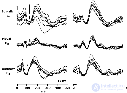
Schematized endogenous components of auditory evoked potentials (B. Rockstroh et al., 1982): a - in response to stimuli relevant to the task; b - the answer to irrelevant stimulus |
Широкое использование метода регистрации ВП стало возможным в результате компьютеризации психофизиологических исследований в 50-60 гг. Первоначально его применение в основном было связано с изучением сенсорных функций человека в норме и при разных видах аномалий. Впоследствии метод стал успешно применяться и для исследования более сложных психических процессов, которые не являются непосредственной реакцией на внешний стимул.
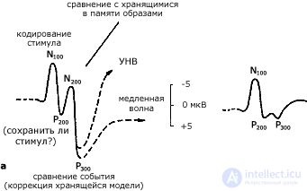
Способы выделения сигнала из шума позволяют отмечать в записи ЭЭГ изменения потенциала, которые достаточно строго связаны во времени с любым фиксированным событием. В связи с этим появилось новое обозначение этого круга физиологических явлений — событийно-связанные потенциалы (ССП).
Эти потенциалы представляют собой последовательность позитивных и негативных колебаний, регистрируемых, как правило, в интервале 0-500 мс. В ряде случаев возможны и более поздние колебания в интервале до 1000 мс. Количественные методы оценки ВП и ССП предусматривают, в первую очередь, оценку амплитуд и латентностей . Амплитуда — размах колебаний компонентов, измеряется в мкВ, латентность — время от начала стимуляции до пика компонента, измеряется в мс. Помимо этого, используются и более сложные варианты анализа.
Феноменологический уровень включает описание ВП как многокомпонентной реакции с анализом конфигурации, компонентного состава и топографических особенностей. Фактически этот уровень анализа, с которого начинается любое исследование, применяющее метод ВП. Возможности этого уровня анализа прямо связаны с совершенствованием способов количественной обработки ВП, которые включают разные приемы, начиная от оценки латентностей и амплитуд и кончая производными, искусственно сконструированными показателями. Многообразен и математический аппарат обработки ВП, включающий факторный, дисперсионный, таксономический и другие виды анализа.
Физиологический уровень. По этим результатам на физиологическом уровне анализа происходит выделение источников генерации компонентов ВП, т.е. решается вопрос о том, в каких структурах мозга возникают отдельные компоненты ВП. Локализация источников генерации ВП позволяет установить роль отдельных корковых и подкорковых образований в происхождении тех или иных компонентов ВП. Наиболее признанным здесь является деление ВП на экзогенные и эндогенные компоненты. Первые отражают активность специфических проводящих путей и зон, вторые — неспецифических ассоциативных проводящих систем мозга. Длительность тех и других оценивается по-разному для разных модальностей. В зрительной системе, например, экзогенные компоненты ВП не превышают 100 мс от момента стимуляции.
Третий уровень анализа — функциональный предполагает использование ВП как инструмента, позволяющего изучать физиологические механизмы поведения и познавательной деятельности человека и животных.
VP as a unit of psychophysiological analysis. The unit of analysis is commonly understood as such an object of analysis, which, unlike the elements, possesses all the basic properties inherent in the whole, and properties are further indecomposable parts of this unity. The unit of analysis is such a minimal formation in which the essential connections and the essential parameters of the object are presented. Moreover, such a unit itself must be a single whole, a kind of system, the further decomposition of which into elements will make it impossible for it to represent the whole as such. An obligatory feature of the unit of analysis is also the fact that it can be operationalized, i.e. it allows measurement and quantitative processing.
If we consider psychophysiological analysis as a method of studying the brain mechanisms of mental activity, then the EAP meet most of the requirements that can be presented to the unit of such analysis.
First , the EP must be qualified as a psychic nervous response, i.e. one that is directly related to the processes of mental reflection.
Secondly , VP is a reaction consisting of a number of components that are continuously interconnected. Thus, it is structurally homogeneous and can be operationalized, i.e. It has quantitative characteristics in the form of parameters of individual components (latencies and amplitudes). It is significant that these parameters have different functional significance depending on the features of the experimental model.
Thirdly , разложение ВП на элементы (компоненты), осуществляемое как метод анализа, позволяет охарактеризовать лишь отдельные стадии процесса переработки информации, при этом утрачивается целостность процесса как такового.
В наиболее выпуклой форме идеи о целостности и системности ВП как корреляте поведенческого акта нашли отражение в исследованиях В.Б. Швыркова. По этой логике ВП, занимая весь временной интервал между стимулом и реакцией, соответствуют всем процессам, приводящим к возникновению поведенческого ответа, при этом конфигурация ВП зависит от характера поведенческого акта и особенностей функциональной системы, обеспечивающей данную форму поведения. При этом отдельные компоненты ВП рассматриваются как отражение этапов афферентного синтеза, принятия решения, включения исполнительных механизмов, достижения полезного результата. В такой интерпретации ВП выступают как единица психофизиологического анализа поведения.
Однако магистральное русло применения ВП в психофизиологии связано с изучением физиологических механизмов и коррелятов познавательной деятельности человека. Это направление определяется как когнитивная психофизиология. ВП в нем используются в качестве полноценной единицы психофизиологического анализа. Такое возможно, потому что, по образному определению одного из психофизиологов, ВП имеют уникальный в своем роде двойной статус, выступая в одно и то же время как "окно в мозг" и "окно в познавательные процессы" (см. Хрестомат. 2.4).
 ТКЭАМ — топографическое картирование электрической активности мозга — область электрофизиологии, оперирующая с множеством количественных методов анализа электроэнцефалограммы и вызванных потенциалов (см. Видео). Широкое применение этого метода стало возможным при появлении относительно недорогих и быстродействующих персональных компьютеров. Топографическое картирование существенным образом повышает эффективность ЭЭГ-метода. ТКЭАМ позволяет очень тонко и дифференцированно анализировать изменения функциональных состояний мозга на локальном уровне в соответствии с видами выполняемой испытуемым психической деятельности. Однако, следует подчеркнуть, что метод картирования мозга является не более чем очень удобной формой представления на экране дисплея статистического анализа ЭЭГ и ВП.
Data logging. The number of electrodes used for recording EEG and EP usually varies in the range from 16 to 32, but in some cases reaches 128 and even more. At the same time, a greater number of electrodes improves the spatial resolution when registering the electric fields of the brain, but it is associated with overcoming great technical difficulties.
To obtain comparable results, the "10-20" system is used, and mainly monopolar registration is used.
It is important that with a large number of active electrodes only one reference electrode can be used, i.e. that electrode with respect to which the EEG of all the other electrode setting points is recorded. The place of application of the reference electrode is the earlobes, nose bridge, or some points on the surface of the scalp (occiput, vertex). There are modifications of this method that make it possible not to use the reference electrode at all, replacing it with the potential values calculated on a computer.
Data analysis. There are several main methods of quantitative analysis of EEG: time, frequency and spatial.
Temporary is a variant of the reflection of EEG and EP data on the graph, while the time is plotted on the horizontal axis, and the amplitude is on the vertical axis. Time analysis is used to estimate the total potentials, peaks of EP, epileptic discharges.
Frequency analysis consists of grouping data by frequency ranges: delta, theta, alpha, beta.
Spatial analysis is associated with the use of various statistical processing methods when comparing EEG from different leads. The most commonly used method is the calculation of coherence.
Ways to present data. The most advanced computer brain mapping tools make it easy to reflect on the display all the stages of analysis: the raw data of the EEG and EP, the power spectra, the topographic maps, both statistical and dynamic in the form of cartoons, various graphs, charts and tables, and to the wish of the researcher, - various complex representations. It should be emphasized that the use of various forms of data visualization allows you to better understand the features of the course of complex brain processes.
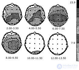 |
EEG-maps representing the topographic location of the spectral power values of the EEG (according to NL Gorbachevskaya et al., 1991). Under each map the range of analyzed frequencies is indicated. Right - the scale of the values of the spectral power of the EEG, µV |
Topographic maps represent the contour of the skull, which shows any color coded EEG parameter at a certain point in time, with different gradations of this parameter (severity) represented by different color shades. Since the EEG parameters are constantly changing as the survey progresses, the color composition on the screen changes accordingly, allowing you to visually monitor the dynamics of the EEG processes. In parallel with the observation, the researcher has at his disposal the statistical data underlying the maps.
The use of TKEA in psychophysiology is most productive when using psychological samples that are "topographically contrasting" , i.e. addressed to different parts of the brain (for example, verbal and spatial assignments).
 Computed tomography (CT) is the newest method that gives accurate and detailed images of the slightest changes in the density of the brain substance. CT has combined the latest achievements of X-ray and computer technology, differing in the fundamental novelty of technical solutions and software.
The main difference between CT and X-ray is that X-rays give only one type of body part. With the help of computed tomography, you can get a lot of images of the same organ and thus build an internal cross section, or "slice" of this part of the body. A tomographic image is the result of accurate measurements and calculations of X-ray attenuation related only to a specific organ.
Thus, the method allows us to distinguish between tissues that differ insignificantly among themselves in terms of their absorptive capacity. The measured radiation and the degree of its attenuation receive a numerical expression. On a set of measurements of each layer, computerized tomogram synthesis is carried out. The final stage is the construction of the image of the investigated layer on the display screen. For tomographic studies of the brain, a neurotomograph is used.
In addition to solving clinical problems (for example, determining the location of a tumor), CT can be used to get an idea of the distribution of regional cerebral blood flow. Thanks to this, CT can be used to study the metabolism and blood supply to the brain.
In the course of life, neurons consume various chemicals that can be labeled with radioactive isotopes (for example, glucose). With the activation of nerve cells, the blood supply to the corresponding part of the brain increases, as a result, labeled substances accumulate in it and radioactivity increases. Measuring the level of radioactivity in different parts of the brain, it is possible to draw conclusions about changes in brain activity in various types of mental activity. Recent studies have shown that determining the most activated areas of the brain can be carried out with an accuracy of 1 mm.
Nuclear magnetic resonance imaging of the brain. Computed tomography has become the parent of a number of other more advanced research methods: tomography using the effect of nuclear magnetic resonance (NMR tomography), positron emission tomography (PET), functional magnetic resonance (FMR). These methods are among the most promising methods of non-invasive combined study of the structure, metabolism and blood flow of the brain.
 In NMR tomography, imaging is based on determining the distribution of the density of hydrogen nuclei (protons) in the medulla and on recording some of their characteristics using powerful electromagnets located around the human body. Images obtained by means of NMR tomography provide information about the studied brain structures of not only anatomical, but also physicochemical nature. In addition, the advantage of nuclear magnetic resonance is the absence of ionizing radiation; in the possibility of multi-plane research carried out exclusively by electronic means; in higher resolution. In other words, using this method, you can get a clear image of "slices" of the brain in different planes.
Positron Emission Transaxial Tomography ( PET scanners ) combines the capabilities of CT and radioisotope diagnostics. It uses ultrashort-lived positron-emitting isotopes ("dyes"), which are part of the natural metabolites of the brain, which are introduced into the human body through the respiratory tract or intravenously. Active brain areas need more blood flow, so more radioactive "dye" accumulates in the working areas of the brain. The radiation of this "dye" is converted into images on the display.
With PET, regional cerebral blood flow and the metabolism of glucose or oxygen in certain parts of the brain are measured. PET allows for intravital mapping on brain slices of regional metabolism and blood flow.
Currently, new technologies are being developed for studying and measuring the processes occurring in the brain, based, in particular, on the combination of the NMR method with the measurement of brain metabolism using positron emission. These technologies are called the functional magnetic resonance (FMR) method (see Video).
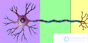 Neuron - the nerve cell through which information is transmitted in the body, is a morphofunctional unit of the central nervous system of humans and animals. When reaching the threshold level of excitation entering the neuron from different sources, it generates a discharge, called the action potential. As a rule, a neuron must receive a lot of incoming pulses before a response discharge occurs in it. All contacts of the neuron ( synapses ) are divided into two classes: excitatory and inhibitory. The activity of the former increases the possibility of discharging a neuron; the activity of the latter decreases it. In figurative comparison, the neuron’s response to the activity of all its synapses is the result of a kind of “chemical voting”. The frequency of neuron responses depends on how often and with what intensity its synaptic contacts are excited, but there are some limitations. The generation of impulses (spikes) makes the neuron incompetent by about 0.001 s. This period is called refractory, it is needed to restore cell resources. The period of refractoriness limits the frequency of neuronal discharges. The frequency of discharges of neurons varies widely, according to some data from 300 to 800 pulses per second (see Video).
 |
Oscillogram variants of impulse activity of neural populations recorded in various cortical and subcortical structures (according to NP Bekhtereva et al., 1985). Above - time stamps (100 ms). Latin letters on the right - symbols of human brain structures |
Registration of neuron responses. The activity of a single neuron is recorded using so-called microelectrodes, the tip of which is from 0.1 to 1 micron in diameter. Special devices allow such electrodes to be introduced into different parts of the brain, in this position the electrodes can be fixed and, when connected to the amplifier-oscilloscope complex, they allow one to observe the electrical discharges of the neuron.
Using microelectrodes, the activity of individual neurons, small ensembles (groups) of neurons, and multiple populations (that is, relatively large groups of neurons) are recorded. The quantitative processing of records of impulse activity of neurons is a rather difficult task, especially in cases where a neuron generates many bits and it is necessary to identify changes in this dynamics depending on some factors. With the help of computers and special software, parameters such as the pulse frequency, the frequency of rhythmic packs or grouping of pulses, the duration of interstimulus intervals, etc. are evaluated. The functional characteristics of neuron activity are analyzed in comparison with behavioral responses for 25–30 s above. The activity of neurons is recorded in animals in the experiment, in humans in a clinical setting. Valuable objects for studying the functional properties of neurons are the large and relatively accessible neurons of some invertebrates. Numerous facts concerning the neuronal organization of behavior were obtained when studying the pulsed activity of neurons in experiments on rabbits, cats and monkeys.
Studies of the activity of human brain neurons are carried out in a clinical setting, when special microelectrodes are introduced into the brain by patients with therapeutic purposes. In the course of treatment, for the completeness of the clinical picture, patients undergo psychological testing, during which the activity of neurons is recorded. The study of bioelectric processes in cells that retain all their connections in the brain, allows you to compare the characteristics of their activity, with the results of psychological tests, on the one hand, as well as with integrative physiological indicators (EEG, VP, EMG, etc.)
The latter is especially important, because one of the tasks of studying the work of the brain is to find such a method that would harmoniously combine the subtlest analysis in studying the details of its work with the study of integral functions. Knowledge of the laws of the functioning of individual neurons, of course, is absolutely necessary, but this is only one side in studying the functioning of the brain, but it does not reveal the laws of how the brain functions as an integral functional system.
Above were presented methods, the general purpose of which is to register the physiological manifestations and indicators of the functioning of the human brain and animals. Along with this, researchers have always sought to penetrate the mechanisms of the brain, exerting direct or indirect effects on it and evaluating the effects of these effects. For the psychophysiologist, the use of various methods of stimulation is a direct possibility of modeling behavior and mental activity in the laboratory.
 Sensory stimulation. The easiest way to influence the brain is to use natural or similar stimuli (visual, auditory, olfactory, tactile, etc.). By manipulating the physical parameters of the stimulus and its substantial characteristics, the researcher can simulate different aspects of mental activity and human behavior.
The range of incentives used is very wide:
in the field of visual perception - from elementary visual stimuli (flashes, chess fields, grids) to visually presented words and sentences, with finely differentiated semantics;
in the field of hearing , from non-speech stimuli (tones, clicks) to phonemes, words and sentences.
In the study of tactile sensitivity, stimulation is applied: mechanical and electrical stimuli that do not reach the pain sensitivity threshold, while irritation can be applied to different parts of the body.
The reactions of the central nervous system to such effects are well studied both by recording the activity of neurons and by the method of evoked potentials. In addition, psychophysiology is widely used methods of rhythmic stimulation by light or sound, causing the effects of imposing - play in the EEG spectrum of frequencies corresponding to the frequency of the current stimulus (or multiples of this frequency).
Electrical brain stimulation is a fruitful method of studying the functions of its individual structures. It is carried out through electrodes introduced into the brain in "acute" animal experiments or during surgical operations on the brain in humans. In addition, stimulation is also possible under long-term observation conditions with the help of electrodes that have been implanted operatively. With chronically implanted electrodes, it is possible to study a special phenomenon of electrical self-stimulation, when an animal closes an electrical circuit with the help of some action (pressing the lever) and thus regulates the force of stimulation of its own brain. In humans, electrical brain stimulation is used to study the relationship between mental processes and functions and parts of the brain. For example, one can study the physiological bases of speech, memory, emotions.
Under laboratory conditions, the method of micropolarization is used, the essence of which consists in passing a weak direct current through separate parts of the cerebral cortex. When this electrode is applied to the surface of the skull in the area of stimulation. Local micropolarization does not destroy brain tissue, but only influences shifts in the potential of the cortex in the stimulated area, so it can be used in psychophysiological studies.
Along with electrical stimulation of the human cerebral cortex by a weak electromagnetic field is permissible. The basis of this method is the fundamental possibility of changing the characteristics of the activity of the central nervous system under the influence of controlled magnetic fields. In this case, there is also no damaging effect on brain cells. At the same time, according to some data, the impact of an electromagnetic field significantly affects the course of mental processes, therefore, this method is of interest to psychophysiology.
The destruction of areas of the brain. Damage or removal of part of the brain to establish its functions in providing behavior is one of the oldest and most common methods for studying the physiological basis of behavior. In its pure form, the method is used in experiments with animals. Along with this, a psychophysiological examination of people who, for medical reasons, were removed part of the brain, is common.
Итак, в общем метод разрушения мозга включает в себя разрушение, удаление и рассечение ткани, истощение нейрохимических веществ, в первую очередь медиаторов, а также временное функциональное выключение отдельных областей головного мозга и оценку влияния вышеперечисленных эффектов на поведение животных.
 Методы регистрации. Измерение и изучение электрической активности кожи (ЭАК), или кожно-гальванической реакции ( КГР ), впервые началось в конце 19 в., когда почти одновременно французский врач Фере и российский физиолог Тарханов зарегистрировали: первый — изменение сопротивления кожи при пропускании через нее слабого тока, второй — разность потенциалов между разными участками кожи. Эти открытия легли в основу двух методов регистрации КГР: экзосоматического (измерение сопротивления кожи) и эндосоматического (измерение электрических потенциалов самой кожи). Следует помнить, что эти методы дают несовпадающие результаты.
 В настоящее время ЭАК объединяет целый ряд показателей: уровень потенциала кожи, реакция потенциала кожи, спонтанная реакция потенциала кожи, уровень сопротивления кожи, реакция сопротивления кожи, спонтанная реакция сопротивления кожи. В качестве индикаторов стали использоваться также характеристики проводимости кожи: уровень, реакция и спонтанная реакция. Во всех трех случаях "уровень" означает тоническую составляющую ЭАК, т.е. длительные изменения показателей; "реакция" — фазическую составляющую ЭАК, т.е. быстрые, ситуативные изменения показателей ЭАК; спонтанные реакции — краткосрочные изменения, не имеющие видимой связи с внешними факторами.
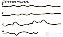
Происхождение и значение ЭАК. Возникновение электрической активности кожи обусловлено, главным образом, активностью потовых желез в коже человека, которые в свою очередь находятся под контролем симпатической нервной системы.
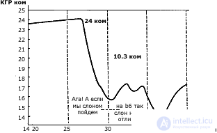 |
Динамика кожно-гальванической реакции в процессе решения мыслительной (шахматной) задачи (по О.К.Тихомирову, 1984). В нижней части рисунка даны сопровождающие решение речевые рассуждения. Резкое падение сопротивления кожи является показателем эмоциональной активации в момент принятия решения |
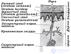
У человека имеется 2-3 миллиона потовых желез, но количество их на разных участках теле сильно варьирует. Например, на ладонях и подошвах около 400 потовых желез на один квадратный сантиметр поверхности кожи, на лбу около 200, на спине около 60. Выделение железами пота происходит постоянно, даже когда на коже не появляется ни капли. В течении дня выделяется около полулитра жидкости. При исключительно сильной жаре потеря жидкости может достигать 3,5 литра в час и 14 литров в день (см. Видео).
Существует два типа потовых желез: апокринные и эккринные .
Апокринные , расположенные в подмышечных впадинах и в паху, определяют запах тела и реагируют на раздражители, вызывающие стресс. Они непосредственно не связаны с регуляцией температуры тела.
 Эккринные расположены по всей поверхности тела и выделяют обычный пот, главными компонентами которого являются вода и хлористый натрий. Их главная функция — терморегуляция, т.е. поддержание постоянной температуры тела. Однако те эккринные железы, которые расположены на ладонях и подошвах ног, а также на лбу и под мышками — реагируют в основном на внешние раздражители и стрессовые воздействия.
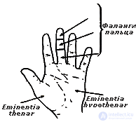
В психофизиологии электрическую активность кожи используют как показатель "эмоционального" потоотделения. Как правило, ее регистрируют с кончиков пальцев или ладони, хотя можно измерять и с подошв ног, и со лба. Следует сказать, однако, что природа КГР , или ЭАК, еще до сих пор не ясна.
Сердечно-сосудистая система выполняет витальные функции, обеспечивая постоянство жизненной среды организма. Сердечная мышца и кровеносные сосуды действуют согласованно, чтобы удовлетворять постоянно меняющиеся потребности различных органов и служить сетью для снабжения и связи, поскольку с кровотоком переносятся питательные вещества, газы, продукты распада, гормоны.
 Среди показателей сердечно-сосудистой системы часто используют также среднюю частоту пульса и ее дисперсию.
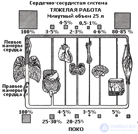
У взрослого человека в состоянии относительного покоя систолический объем каждого желудочка составляет 70-80 мл. Минутный объем сердца — количество крови, которое сердце выбрасывает в легочный ствол и аорту за 1 мин — измеряется как произведение величины систолического объема на частоту сердечных сокращений в 1 мин. В покое минутный объем составляет 3-5 л. При интенсивной работе минутный объем может существенно увеличиваться до 25-30 л., причем на первых этапах минутный объем сердца растет за счет повышения величины систолического объема, а при больших нагрузках в основном за счет увеличения сердечного ритма.
Артериальное давление — общеизвестный показатель работы сердечно-сосудистой системы. Оно характеризует силу напора крови в артериях. АД изменяется на протяжении сердечного цикла, оно достигает максимума во время систолы (сокращения сердца) и падает до минимума в диастоле, когда сердце расслабляется перед следующим сокращением. Нормальное артериальное давление здорового человека в покое около 130 / 70 мм рт.ст., где 130 — систолическое давление АД, а 70 — диастолическое АД. Пульсовое давление разность между систолическим и диастолическим давлением, и в норме составляет около 60 мм рт.ст.
Ритм сердца — показатель, часто используемый для диагностики функционального состояния человека, зависит от взаимодействия симпатических и парасимпатических влияний из вегетативной нервной системы. При этом возрастание напряженности в работе сердца может возникать по двум причинам — в результате усиления симпатической активности и снижения парасимпатической.
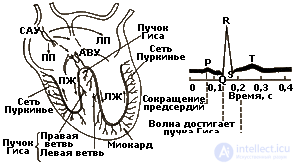
Электрокардиограмма (ЭКГ) — запись электрических процессов, связанных с сокращением сердечной мышцы . Впервые была сделана в 1903 г. Эйнтховеном. С помощью клинических и диагностических установок ЭКГ можно регистрировать, используя до 12 различных пар отведений; половина их связана с грудной клеткой, а другая половина — с конечностями. Каждая пара электродов регистрирует разность потенциалов между двумя сторонами сердца, и разные пары дают несколько различную информацию о положении сердца в грудной клетке и о механизмах его сокращения. При заболеваниях сердца в одном или нескольких отведениях могут обнаруживаться отклонения от нормальной формы ЭКГ, и это существенно помогает при постановке диагноза.
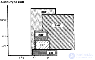 |
Амплитудно-частотные соотношения биоэлектрических сигналов (ЭЭГ, ЭМГ, ЭОГ, ЭКГ) (по В.В. Гнездицкому, 1997) |
 В психофизиологии ЭКГ в основном используется для измерения частоты сокращения желудочков. С этой целью применяют прибор кардиотахометр. Ритм сердца, зарегистрированный с помощью кардиотахометра, как правило, соответствует частоте пульса, т.е. числу волн давления, распространяющихся вдоль периферических артерий за одну минуту. В некоторых случаях эти величины, однако, не совпадают.
 Исследование нейрогуморальной регуляции ритма сердца является одним из наиболее распространенных подходов к оценке состояния адаптационных возможностей организма человека. Для исследования вегетативного тонуса широко используются записи ЭКГ или кардиоинтервалограммы (КИГ). Наиболее распространенным является метод обработки кардиоинтервалов с помощью гистографического анализа: вычисляется мода распределения, ее амплитуда и вариационный размах и на основании этих параметров вычислялся интегральный показатель — индекс напряжения (ИН). Индекс напряжения пропорционален средней частоте сердечных сокращений и обратно пропорционален диапазону, в котором варьирует интервал между двумя ударами сердца.
С начала 60-х гг. начали использоваться различные спектральные методы анализа RR-интервалов.
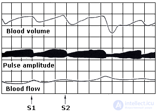 Плетизмография — метод регистрации сосудистых реакций организма . Плетизмография отражает изменения в объеме конечности или органа, вызванные изменениями количества находящейся в них крови. Конечность человека в изолирующей перчатке помещают внутрь сосуда с жидкостью, который соединен с манометром и регистрирующим устройством. Изменения давления крови и лимфы в конечности находят отражение в форме кривой, которая называется плетизмограммой. Широкое распространение получили пальцевые фотоплетизмографы, портативные устройства, которые также можно использовать для регистрации сердечного ритма.
 В плетизмограмме можно выделить два типа изменений: фазические и тонические.
Фазические изменения обусловлены динамикой пульсового объема от одного сокращения сердца к другому.
Тонические изменения кровотока — это собственно изменения объема крови в конечности. Оба показателя обнаруживают при действии психических раздражителей сдвиги, свидетельствующие о сужении сосудов.
Плетизмограмма — высоко чувствительный индикатор вегетативных сдвигов в организме.
Мышечную систему образно определяют как биологический ключ человека к внешнему миру.
 Электромиография — метод исследования функционального состояния органов движения путем регистрации биопотенциалов мышц . Электромиография — это регистрация электрических процессов в мышцах, фактически запись потенциалов действия мышечных волокон, которые заставляют ее сокращаться. Мышца представляет собой массу ткани, состоящую из множества отдельных мышечных волокон, соединенных вместе и работающих согласованно. Каждое мышечное волокно — это тонкая нить, толщиной всего лишь около 0,1 мм до 300 мм длиной. При стимуляции электрическим потенциалом действия, приходящим к волокну от мотонейрона, это волокно сокращается иногда примерно до половины первоначальной длины. Мышцы, участвующие в тонких двигательных коррекциях (фиксация объекта глазами), могут иметь в каждой единице всего по 10 волокон. В мышцах, осуществляющих более грубую регулировку при поддержании позы, в одной двигательной единице может быть до 3000 мышечных волокон.
 Поверхностная электромиограмма (ЭМГ) суммарно отражает разряды двигательных единиц, вызывающих сокращение. Регистрация ЭМГ позволяет выявить намерение начать движение за несколько секунд до его реального начала. Помимо этого миограмма выступает как индикатор мышечного напряжения. В состоянии относительного покоя связь между действительной силой, развиваемой мышцей, и ЭМГ линейна.
Прибор, с помощью которого регистрируются биопотенциалы мышц, называется электромиографом, а регистрируемая с его помощью запись электромиограммой (ЭМГ). ЭМГ, в отличие от биоэлектрической активности мозга (ЭЭГ), состоит из высокочастотных разрядов мышечных волокон, для неискаженной записи которых, по некоторым представлениям, требуется полоса пропускания до 10 000 Гц.
 Дыхательная система состоит из дыхательных путей и легких.
Основной двигательный аппарат этой системы составляют межреберные мышцы, диафрагма и мышцы живота. Воздух, поступающий в легкие во время вдоха, снабжает протекающую по легочным капиллярам кровь кислородом. Одновременно из крови выходят двуокись углерода и другие вредные продукты метаболизма, которые выводятся наружу при выдохе. Между интенсивностью мышечной работы, совершаемой человеком, и потреблением кислорода существует простая линейная зависимость.
В психофизиологических экспериментах в настоящее время дыхание регистрируется относительно редко, главными образом для того, чтобы контролировать артефакты.
 Для измерения интенсивности (амплитуды и частоты) дыхания используют специальный прибор — пневмограф. Он состоит из надувной камеры-пояса, плотно оборачиваемой вокруг грудной клетки испытуемого, и отводящей трубки, соединенной с манометром и регистрирующим устройством. Возможны и другие способы регистрации дыхательных движений, но в любом случае обязательно должны присутствовать датчики натяжения, фиксирующие изменение объема грудной клетки.
Этот метод обеспечивает хорошую запись изменений частоты и амплитуды дыхания. По такой записи легко анализировать число вдохов в минуту, а также амплитуду дыхательных движений в разных условиях. Можно сказать, что дыхание — это один из недостаточно оцененных факторов в психофизиологических исследованиях.
Для психофизиолога наибольший интерес представляют три категории глазных реакций: сужение и расширение зрачка, мигание и глазные движения.
Пупиллометрия — метод изучения зрачковых реакций. Зрачок — отверстие в радужной оболочке, через которое свет попадает на сетчатку. Диаметр зрачка человека может меняться в пределах от 1,5 до 9 мм. Величина зрачка существенно колеблется в зависимости от количества света, падающего на глаз: на свету зрачок сужается, в темноте — расширяется. Наряду с этим, размер зрачка существенно изменяется, если испытуемый реагирует на воздействие эмоционально. В связи с этим пупиллометрия используется для изучения субъективного отношения людей к тем или иным внешним раздражителям.
Диаметр зрачка можно измерять путем простого фотографирования глаза в ходе обследования или же с помощью специальных устройств, преобразующих величину зрачка в постоянно варьирующий уровень потенциала, регистрируемый на полиграфе.
Мигание (моргание) — периодическое смыкание век . Длительность одного мигания приблизительно 0,35 с. Средняя частота мигания составляет 7,5 в минуту и может варьировать в пределах от 1 до 46 в минуту. Мигание выполняет разные функции в обеспечении жизнедеятельности глаз. Однако для психофизиолога существенно, что частота мигания изменяется в зависимости от психического состояния человека.
Движение глаз широко исследуются в психологии и психофизиологии. Это разнообразные по функции, механизму и биомеханике вращения глаз в орбитах. Существуют разные типы глазных движений, выполняющие различные функции. Однако наиболее важная среди них функция движений глаз состоит в том, чтобы поддерживать интересующее человека изображение в центре сетчатки, где самая высокая острота зрения. Минимальная скорость прослеживающих движений около 5 угл. мин/с, максимальная достигает 40 град/с.
Электроокулография — метод регистрации движения глаз , основанный на графической регистрации изменения электрического потенциала сетчатки и глазных мышц. У человека передний полюс глаза электрически положителен, а задний отрицателен, поэтому существует разность потенциалов между дном глаза и роговицей, которую можно измерить. При повороте глаза положение полюсов меняется, возникающая при этом разность потенциалов характеризует направление, амплитуду и скорость движения глаза. Это изменение, зарегистрированное графически, носит название электроокулограммы. Однако микродвижения глаз с помощью этого метода не регистрируются, для их регистрации разработаны другие приемы. (см. рис.)
 Детектор лжи — условное название прибора полиграфа, одновременно регистрирующего комплекс физиологических показателей ( КГР , ЭЭГ, плетизмограмму и др.) с целью выявить динамику эмоционального напряжения. С человеком, проходящем обследование на полиграфе, проводят собеседование, в ходе которого наряду с нейтральными задают вопросы, составляющие предмет специальной заинтересованности. По характеру физиологических реакций, сопровождающих ответы на разные вопросы, можно судить об эмоциональной реактивности человека и в какой-то мере о степени его искренности в данной ситуации. Поскольку в большинстве случаев специально необученный человек не контролирует свои вегетативные реакции, детектор лжи дает по некоторым оценкам до 71% случаев обнаружения обмана.
Следует иметь в виду, однако, что сама процедура собеседования (допроса) может быть настолько неприятна для человека, что возникающие по ходу физиологические сдвиги будут отражать эмоциональную реакцию человека на процедуру. Отличить спровоцированные процедурой тестирования эмоции от эмоций, вызванных целевыми вопросам, невозможно. В то же время человек, обладающий высокой эмоциональной стабильностью, сможет относительно спокойно чувствовать себя в этой ситуации, и его вегетативные реакции не дадут твердых основания для вынесения однозначного суждения. По этой причине к результатам, полученных с помощью детектора лжи, нужно относиться с должной мерой критичности (см. Видео).
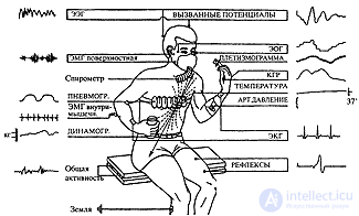 |
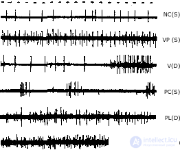
| Многоканальная регистрация наиболее часто изучаемых видов биоэлектрической активности человека (по В.Блоку, 1970) |
В идеале выбор физиологических методик и показателей должен логически вытекать из принятого исследователем методологического подхода и целей, поставленных перед экспериментом. Однако на практике нередко исходят из других соображений, например, доступности приборов и легкости обработки экспериментальных данных.
Более весомыми представляются аргументы в пользу выбора методик, если извлекаемые с их помощью показатели получают логически непротиворечивое содержательное толкование в контексте изучаемой психологической или психофизиологической модели.
Psychophysiological models. In science, a model is understood as simplified knowledge, carrying certain, limited information about an object / phenomenon, reflecting some of its properties. With the help of models, it is possible to imitate the functioning and predict the properties of the objects, processes or phenomena being studied. In psychology, modeling has two aspects: modeling the psyche and modeling situations . The first implies a sign or technical imitation of the mechanisms, processes and results of mental activity, the second is the organization of a particular type of human activity by artificially constructing the environment in which this activity takes place.
Both aspects of modeling find a place in psycho-physiological research. In the first case, the simulated features of human activity, mental processes and states are predicted on the basis of objective physiological parameters, often recorded outside of a direct connection with the phenomenon under study. For example, it is shown that some of the individual characteristics of perception and memory can be predicted by the characteristics of the brain biocurrents. In the second case, psychophysiological modeling includes imitation in laboratory conditions of a certain mental activity in order to identify its physiological correlates and / or mechanisms. It is obligatory to create some artificial situations in which the investigated mental processes and functions are involved in one way or another. An example of this approach is the numerous experiments to identify the physiological correlates of perception, memory, etc.
When interpreting the results in such experiments, the researcher must clearly understand that the model is never completely identical to the phenomenon or process under study. As a rule, it takes into account only some separate aspects of reality. Consequently, no matter how exhaustive, for example, any psychophysiological experiment in identifying the neurophysiological correlates of memory processes, it will provide only partial knowledge of the nature of its physiological mechanisms, limited by the framework of this model and the methodological techniques and indicators used. It is for this reason that psychophysiology is replete with a variety of unrelated, and sometimes simply contradictory experimental data. Obtained in the context of different models, such data represent fragmentary knowledge, which in the future is likely to unite into an integral system describing the mechanisms of psychophysiological functioning.
Interpretation of indicators. The question of what importance the experimenter attaches to each of the indicators he uses deserves special attention. In principle, physiological indicators can fulfill two main roles: target (sense) and service (auxiliary). For example, when studying the brain biocurrents in the process of mental activity, it is advisable to simultaneously record eye movements, muscle tension, and some other indicators. Moreover, in the context of such work, only indicators of the brain's biocurrents carry the meaning associated with this task. The remaining indicators are used to control artifacts and the quality of registration of biocurrents (registration of eye movements), control of the emotional state of the subject (registration of GSR ), since it is well known that eye movements and emotional stress can introduce interference and distort the pattern of biocurrents. either task. At the same time, in another study, registration and eye movements, and KGR can play a semantic, rather than an official role. For example, when the subject of research is a strategy of visual search or the study of the physiological mechanisms of a person’s emotional sphere.
Thus, the same physiological indicator can be used to solve different problems. In other words, the specific use of an indicator is determined not only by its own functional capabilities, but also by the psychological context in which it is included. Good knowledge of the nature and all the possibilities of the physiological parameters used is an important factor in the organization of the psychophysiological experiment.
The value of experiments performed on animals. As noted above, many problems in psychophysiology were solved and continue to be solved in animal experiments. (First of all, we are talking about the study of the activity of neurons.) In this connection, the problem formulated by LS is of particular importance. Vygotsky. This is the problem of the human-specific ratio of structural and functional units in brain activity and the definition of new, compared to animals, principles for the functioning of systems, intra- and intersystem interactions.
It should be directly pointed out that the problem of the “human-specific ratio of structural and functional units in brain activity and the definition of new compared to animals” principles of the functioning of systems, unfortunately, has not yet received a productive development. According to OS Andrianov (1993): The "rapid" immersion "of biology and medicine ... in the depths of living matter pushed into the background the study of the most important problem - the evolutionary specifics of the human brain. Attempts to find at the molecular level a certain material substrate characteristic only of the human brain and defining features most complex mental functions until they were successful. "
Thus, the question arises about the legality of the transfer of data obtained on animals to explain the brain functions in humans. The point of view is widely accepted, according to which there are universal mechanisms of cellular functioning and general principles of coding information, which allows interpolation of results (see, for example: Fundamentals of Psychophysiology, edited by YI Alexandrova, 1998).
One of the founders of the national psychophysiology E.N. Sokolov, solving the problem of transferring the results of studies performed on animals to humans, formulated the principle of psychophysiological research as follows: man - neuron - model. This means that psychophysiological research begins with the study of human behavioral (psychophysiological) reactions, then it proceeds to the study of behavior mechanisms using microelectrode recording of neural activity in animal experiments, and in humans using electroencephalogram and evoked potentials. The integration of all data is carried out by building a model of neural-like elements. At the same time, the whole model as a whole should reproduce the function under study, and individual neural-like elements should possess the characteristics and properties of real neurons. Prospects for this kind of research lie in building models of a "specifically human type" such as neurointelligence, for example.
Conclusion The above materials indicate a wide variety and different levels of psycho-physiological methods. Much goes into the competence of the psychophysiologist, ranging from the dynamics of neuronal activity in the deep structures of the brain to local blood flow in the finger. The question naturally arises how to combine such different in terms of production methods and content indicators into a logically consistent system. Its solution, however, rests in the absence of a single generally accepted psychophysiological theory.
Psychophysiology, which was born as an experimental branch of psychology, largely remains so to this day, compensating for the imperfection of the theoretical foundation with the diversity and sophistication of the methodical arsenal. The wealth of this arsenal is great, its resources and prospects seem inexhaustible. The rapid growth of new technologies will inevitably expand the possibilities for penetrating the secrets of human physicality. It will lead to the creation of new processing devices capable of formalizing a complex system of dependencies of variables used in objective physiological indicators, naturally associated with human mental activity. Regardless of whether new decisions will result in the further development of computer technology, heuristic models or other methods of knowledge that are not yet known to us, the development of science in our time anticipates a fundamental transformation of psychophysiological thinking and working methods.
1.Is it true that there are three fundamentally different approaches to the analysis of EEG: auditory, clinical and statistical?
1.0
2.Is it true that the evoked potentials are divided into exogenous, mesogenic and endogenous components?
1.0,
3. Is it a judgment that an increase in tension in the work of the heart may arise as a result of a decrease in sympathetic activity?
1.0,
4. Choose the correct answer. H. Berger discovered that brain waves can be recorded from the surface of the skull in:
0.1.0.0,
5. Choose the correct answer. The basic rhythm of an encephalogram of a healthy adult with eyes closed is:
0,0,1,0,
6. Choose the correct answer. Evoked Potentials (EF) - bioelectric oscillations arising in the nervous structures:
0.1.0.0,
7. Choose the correct answer. The evoked potentials are a sequence of positive and negative fluctuations recorded in the interval:
1,0,0,0,
8. Choose the correct answer. The main difference between computed tomography and X-ray is that:"
0,0,0,1,
9. Choose three correct answers. New methods of computed tomography include:
1,1,0,1,
10. Choose two correct answers. All neuron contacts are divided into several classes:
1,0,0,0,
11. Choose the correct answer. The generation of pulses makes a neuron approximately incapable
0.1.0.0,
12. Choose the correct answer. The electrical conductivity of the skin is caused by the condition:
1,0,0,0,
13. Choose the correct answer. The normal blood pressure of a healthy person at rest is:
0.1.0.0,
14. Choose the correct answer. The electrocardiogram was first made by Einthoven at:
1,0,0,0,
15. Choose the correct answer. A cardiotachometer serves in psychophysiology for measuring ECG:
0.1.0.0,
16. Choose the correct answer. Registering an electromyogram reveals an intention to start a movement:
0,1,0,
17. Choose the correct answer. The lie detector provides information:
0,0,1,0,
18. Choose the correct answer. The positron emission tomography method allows you to study:
1,0,0,

Comments
To leave a comment
Psychophysiology
Terms: Psychophysiology