Lecture
Sensory system (analyzer) - is called the part of the nervous system consisting of perceiving elements - sensory receptors, nerve pathways that transmit information from receptors to the brain and parts of the brain that process and analyze this information
The sensor system consists of 3 parts.
1. Receptors - sense organs
2. Conductor-receptor binding to the brain
3. The division of the cerebral cortex, which perceives and processes information.
Receptors are a peripheral link intended for the perception of stimuli of the external or internal environment.
Sensory systems have a general structure plan and are characteristic of sensory systems.
Multi -layering - the presence of several layers of nerve cells, the first of which is associated with receptors, and the latter with neurons of the motor regions of the cerebral cortex. Neurons are specialized for processing various types of sensory information.
Multi - channel - the presence of multiple parallel channels of processing and transmitting information, which provides detailed analysis of signals and greater reliability.
A different number of elements in adjacent layers , which forms the so-called “sensory funnels” (tapering or expanding) They can ensure the elimination of redundancy of information or, conversely, fractional and complex analysis of the signs of a signal
Differentiation of the sensory system vertically and horizontally. Differentiation in the vertical direction means the formation of parts of the sensory system consisting of several neural layers (olfactory bulbs, cochlear nuclei, articulated bodies).
Horizontal differentiation represents the presence of receptors and neurons of different properties within a single layer. For example, the rods and cones in the retina process information in different ways.
The main task of the sensory system is the perception and analysis of the properties of stimuli, on the basis of which sensations, perceptions, and ideas arise. It is a form of sensual, subjective reflection of the outside world.
Functions of sensory systems
Sensory systems are able to respond to inadequate stimuli. If you try the battery terminals, it causes a taste sensation - sour, this is the action of an electric current. Such a reaction of the sensory system to adequate and inadequate stimuli put the question to the physiology - how much can we trust our senses.
Johann Muller formulated in 1840 the law of the specific energy of the sense organs.
The quality of sensations does not depend on the nature of the stimulus, but is determined entirely by the specific energy inherent in the sensitive system, which is released under the action of the stimulus.
With this approach, we can only know what is inherent in ourselves, and not that in the surrounding world. Subsequent studies have shown that excitations in any sensory system arise on the basis of a single energy source, ATP.
A pupil of Muller, Helmholtz, created the theory of symbols , in accordance with which he regarded sensations as symbols and objects of the surrounding world. The theory of symbols denied the possibility of knowing the world around us.
These 2 directions were called physiological idealism. What is the feeling? Sensation is a subjective image of the objective world. Sensations are images of the outside world. They exist in us and are generated by the action of things on our senses. For each of us, this image will be subjective, i.e. it depends on the degree of our development, experience, and each person perceives the surrounding objects and phenomena in their own way. They will be objective, i.e. it means that they exist, regardless of our consciousness. Since there is a subjectivity of perception, then how to decide who most correctly perceives? Where is the truth? The criterion of truth is practical activity. There is a consistent knowledge. At each stage, obtained new information. The child tastes toys, parses them into details. It is on the basis of this deep experience that we acquire deeper knowledge of the world.
Receptor classification.
By the nature of information perception
Receptor arousal mechanisms
In the case of primary receptors, the action of the stimulus is perceived by the end of the sensory neuron. An active stimulus can cause hyperpolarization or depolarization of the surface membrane receptors mainly due to changes in sodium permeability. An increase in permeability to sodium ions leads to membrane depolarization and a receptor potential arises on the receptor membrane. It exists as long as the stimulus acts.
The receptor potential does not obey the law "All or nothing", its amplitude depends on the strength of the stimulus. He has no period of refractoriness. This allows one to sum up the receptor potentials under the action of subsequent stimuli. It spreads meleno, with fading. When the receptor potential reaches a critical threshold, it causes the appearance of an action potential in the nearest interception of Ranvier. In the interception of Ranvier, an action potential arises that obeys the law “All or nothing”. This potential will be pervasive.
In the secondary receptor, the action of the stimulus is perceived by the receptor cell. In this cell, a receptor potential arises, the consequence of which will be the isolation of the mediator from the cell into the synapse, which acts on the postsynaptic membrane of the sensitive fiber and the interaction of the mediator with the receptors leads to the formation of another, local potential, which is called generating . It is identical in properties to receptor. Its amplitude is determined by the amount of the mediator released. Mediators - acetylcholine, glutamate.
Action potentials occur periodically, because they are characterized by a period of refractoriness, when the membrane loses the property of excitability. Action potentials arise discretely and the receptor in the sensory system works as an analog-discrete transducer. At the receptors, adaptation is observed - adaptation to the action of stimuli. There are fast-adapting, there are slowly adapting. When adapting, the amplitude of the receptor potential and the number of nerve impulses that go along the sensitive fiber are reduced. Receptors encode information. It is possible by the frequency of potentials, by the grouping of pulses into separate volleys and the intervals between volleys. Coding is possible by the number of activated receptors in the receptive field.
The threshold of irritation and the threshold of entertainment.
The threshold of irritation is the minimum strength of the stimulus that causes the sensation.
Threshold entertainment - the minimum force of change of the stimulus, at which a new sensation arises.
Hair cells are excited when hairs are displaced by 10 to -11 meters - 0.1 amstrom.
In 1934, Weber formulated a law establishing the relationship between the initial force of irritation and the intensity of sensation. He showed that the change in the strength of the stimulus, is constant.
∆I / Io = К Io = 50 ∆I = 52.11 Io = 100 ∆I = 104.2
Fechner determined that the sensation is directly proportional to the logarithm of stimulation
S = a * logR + b S-feeling R-irritation
S = KI in A Degree I - the strength of irritation, K and A - constants
For tactile receptors, S = 9.4 * I d 0.52
Sensory systems have receptors for self-regulation of receptor sensitivity.
The influence of the sympathetic system - the sympathetic system increases the sensitivity of the receptors to the action of stimuli. This is useful in a danger situation. Increases the excitability of receptors - reticular formation. As part of the sensory nerves, efferent fibers have been found that can alter the sensitivity of the receptors. Such nerve fibers are in the auditory organ.
Sensory hearing system
For most people living in a modern halt, their hearing progressively drops. This happens with age. This is due to environmental pollution - motor vehicles, a disco, etc. The changes in the hearing aid are not reversible. Human ears contain 2 sensitive organs. Hearing and balance. Sound waves propagate in the form of compressions and discharges in elastic media, and the propagation of sounds in dense media is better than in gases. The sound has 3 important properties - height or frequency, power, or intensity and timbre. The pitch of the sound depends on the frequency of vibrations and perceives the human ear with a frequency of 16 to 20,000 Hz. With a maximum sensitivity of 1000 about 4000 Hz.
The main frequency of the sound of the larynx of a man is 100 Hz. Women - 150 Hz. When talking, there are additional high-frequency sounds in the form of hissing, whistling, which disappear when talking on the phone and this makes the speech clearer.
Sound power is determined by the amplitude of oscillations. Sound power is expressed in dB. Power is a logarithmic relationship. Whispered speech - 30 dB, normal speech - 60-70 dB. The sound of transport - 80, the noise of the engine of the aircraft - 160. The sound power of 120 dB causes discomfort, and 140 lead to painful sensations.
The timbre is determined by the secondary oscillations on the sound waves. Ordered vibrations - create musical sounds. And random fluctuations cause just noise. The same note sounds differently on different instruments due to various additional fluctuations.
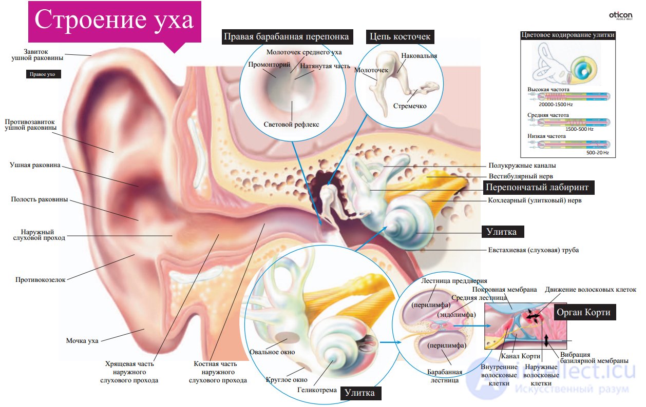
The human ear has 3 component parts - the outer, middle and inner ear. The outer ear is represented by the auricle, which acts as a sound catch funnel. A man’s ear is less completely catching sounds than a rabbit, a horse that knows how to control its ears. At the base of the auricle is cartilage, with the exception of the earlobe. Cartilage tissue gives elasticity and shape to the ear. If the cartilage is damaged, then it regenerates growing. The external auditory canal is S-shaped — inward, forward and down, 2.5 cm long. The auditory canal is covered with skin with low sensitivity of the external part and high sensitivity of the internal. In the outer part of the ear canal there are hairs that prevent particles from entering the ear canal. The glands of the ear canal produce a yellow lubricant that also protects the ear canal. At the end of the aisle, there is a tympanic membrane, which consists of fibrous fibers, covered outside by the skin, and inside - by the mucous membrane. The eardrum separates the middle from the outer ear. It oscillates with the frequency of the perceived sound.
The middle ear is represented by the tympanic cavity, the volume of which is about 5-6 drops of water and the tympanic cavity is filled with water, lined with mucous membranes and contains 3 auditory ossicles: the malleus, anvil and stirrup. At rest, the lumen of the Eustachian tube is closed, which equalizes the pressure. Inflammatory processes that lead to inflammation of this tube cause a feeling of congestion. The middle ear is separated from the inner oval and round hole. Oscillations of the eardrum through the system of levers are transmitted by a stirrup to the oval window, with the outer ear transmitting sounds by air.
There is a difference in the area of the tympanic membrane and the oval window (the area of the eardrum is 70 mm in sq. And the oval window is 3.2 mm in sq.). When the oscillation is transferred from the membrane to the oval window, the amplitude decreases and the oscillation force increases 20-22 times. At frequencies up to 3000 Hz, 60% of E is transmitted to the inner ear. In the middle ear, there are 2 muscles that alter oscillations: a muscle straining the eardrum (attached to the central part of the eardrum and to the handle of the malleus) - with an increase in the force of contraction, the amplitude decreases; the stirrup muscle; its contractions limit the stirrup fluctuations. These muscles prevent injury to the eardrum. In addition to the air transmission of sounds, there is also a bone transmission, but this sound power is not able to cause oscillations of the skull bones.
Inside ear
The inner ear is a maze of interconnected tubules and extensions. In the inner ear there is an organ of balance. The labyrinth has a bone base, and inside there is a webbed labyrinth and there is an endolymph. The cochlea belongs to the auditory part, it forms 2.5 turns around the central axis and is divided into 3 ladders: vestibular, drum and webbed. The vestibular canal begins with an oval window membrane, and ends with a round window. At the top of the cochlea, these 2 channels communicate with the help of the helioccreme. And both of these channels are filled with perilymph. On average, the membranous canal is a sound-perceiving apparatus — the organ of Corti. The main membrane is constructed of elastic fibers that start at the base (0.04mm) and to the top (0.5mm). Towards the top, the fiber density decreases 500 times. On the main membrane is the organ of Corti. It is built from 20-25 thousand special hair cells located on supporting cells. The hair cells lie in 3-4 rows (outer row) and in one row (inner). At the top of the hair cells there are stereocili or kinocili, the largest stereocili. Sensory fibers of 8 pairs of FMN from the spiral ganglion are suitable for hair cells. At the same time 90% of the selected sensitive fibers are on the internal hair cells. On one internal hair cell converges up to 10 fibers. And in the composition of the nerve fibers there are efferent (olive-snail bundle). They form inhibitory synapses on the sensory fibers of the spiral ganglion and innervate the outer hair cells. Irritation of the organ of Corti is associated with the transfer of oscillations of the bones on the oval window. Low-frequency vibrations propagate from the oval window to the top of the cochlea (the entire main membrane is involved). At low frequencies, excitation of the hair cells lying on the top of the cochlea is observed. Bekashi was studying the propagation of waves in the cochlea. He found that with increasing frequency, a smaller column of fluid was involved. Высокочастотные звуки не могут вовлечь весь столб жидкости, поэтому чем больше частота, тем меньше колеблется перилимфа. Колебания основной мембраны могут возникать при передаче звуков через перепончатый канал. При колебании основной мембраны происходит смещение волосковых клеток вверх, что вызывает деполяризацию, а если вниз- волоски отклоняются внутрь, что приводит к гиперполяризации клеток. При деполяризации волосковых клеток открываются Са-каналы и Са способствует потенциалу действия, который несет информацию о звуке. Наружные слуховые клетки имеют эфферентную иннервацию и передача возбуждения идет с помощью Асh на наружных волосковых клетках. Эти клетки могут изменять свою длину: они укорачиваются при гиперполяризации и удлиняются при поляризации. Изменение длины наружных волосковых клеток влияет на колебательный процесс, что улучшает восприятие звука внутренними волосковыми клетками. Изменение потенциала волосковых клеток связано с ионным составом эндо- и перилимфы. Перилимфа напоминает ликвор, а эндолимфа имеет высокую концентрацию К(150 ммоль). Поэтому эндолимфа приобретает положительный заряд к перилифме.( +80мВ). Волосковые клетки содержат много К; они имеют мембранный потенциал и отрицательно заряженный внутри и положительный снаружи(МП=-70мВ), а разница потенциалов дает возможность проникновения К из эндолимфы внутрь волосковых клеток. Изменение положения одного волоска открывает 200-300 К- каналов и возникает деполяризация. Закрытие сопровождается гиперполяризацией. В кортиевом органе идет частотное кодирование за счет возбуждения разных участков основной мембраны. При этом было показано что звуки низкой частоты могут кодироваться числом нервных импульсов таким же количеством как и звуком. Такое кодирование возможно при восприятии звука до 500Гц. Кодирование информации звука достигается увеличением числа залпов волокон на более интенсивный звук и за счет числа активирующихся нервных волокон. Чувствительные волокна спирального ганглия оканичиваются в дорсальных и вентральных ядрах улитки продолговатого мозга. От этих ядер сигнал поступает в ядра оливы как своей так и противоположной стороны. От ее нейронов идут восходящие пути в составе латеральной петли которые подходят к нижним бугоркам четверохолмия и медиальному коленчатому телу зрительного бугра. От последнего сигнал идет в верхнюю височную извилину( извилина Гешля). Это соответствует 41 и 42 полям( первичная зона) и 22 поле( вторичная зона). В ЦНС существует топотоническая организация нейронов, то есть воспринимаются звуки с разной частотой и разной интенсивностью. Корковый центр имеет значение для восприятия, последовательности звука и пространственной локализации. При поражении 22 поля нарушается определение слов (рецептивная оппозия).
Ядра верхней оливы делят на медиальные и латеральные части. А латеральные ядра определяют неодинаковую интенсивность звуков, поступающих к обеим ушам. Медиальное ядро верхней оливы улавливает временные различия поступления звуковых сигналов. Обнаружено что сигналы от обоих ушей поступают в различные дендритные системы одного и того же воспринимающего нейрона. Нарушение слухового восприятия может проявляться звоном в ушах при раздражении внутреннего уха или слухового нерва и двумя типами глухоты: проводниковой и нервной. Первая связана с поражениями наружного и среднего уха( серная пробка).Вторая связана с дефектами внутреннего уха и поражениями слухового нерва. У пожилых людей утрачивается способность воспринимать высокочастотные голоса. За счет двух ушей можно определять пространственную локализацию звука. Это оказывается возможным, если звук отклоняется от средины положения на 3 градуса. При восприятии звуков возможно развитие адаптации за счет ретикулярной формации и эфферентных волокон( воздействием на наружные волосковые клетки.
Зрительная система.
Зрение – многозвеньевой процесс, начинающийся с проекции изображения на сетчатку глаза, затем идёт возбуждение фоторецепторов, передача и преобразование в нейронных слоях зрительной системы и заканчивается принятием высшими корковыми отделами решения о зрительном образе.
Строение и функции оптического аппарата глаза. Глаз имеет шарообразную форму, что важно для поворота глаза. Свет проходит через несколько прозрачных сред – роговицу, хрусталик и стекловидное тело, имеющие определённые преломляющие силы, выражающихся в диоптриях. Диоптрия равна преломляющей силе линзы с фокусным расстоянием 100 см. Преломляющая сила глаза при рассматривании далёких предметов – 59D, близких – 70,5D. На сетчатке образуется уменьшенное перевёрнутое изображение.
Аккомодация – приспособление глаза к ясному видению предметов на разных расстояниях. Хрусталик играет главную роль в аккомодации. При рассмотрении близких предметов ресничные мышцы сокращаются, циннова связка расслабляется, хрусталик становится более выпуклым в силу его эластичности. При рассмотрении дальних – мышцы расслаблены, связки натянуты и растягивают хрусталик, делая его более уплощённым. Ресничные мышцы иннервируются парасимпатическими волокнами глазодвигательного нерва. В норме дальняя точка ясного видения – в бесконечности, ближайшая – 10 см от глаза. Хрусталик с возрастом теряет эластичность, поэтому ближайшая точка ясного видения отодвигается и развивается старческая дальнозоркость.
Аномалии рефракции глаза.
Близорукость (миопия). Если продольная ось глаза слишком длинная или увеличивается преломляющая сила хрусталика, то изображение фокусируется перед сетчаткой. Человек плохо видит вдаль. Назначаются очки с вогнутыми стёклами.
Дальнозоркость (гиперметропия). Развивается при уменьшении преломляющих сред глаза или при укорочении продольной оси глаза. В результате изображение фокусируется за сетчаткой и чел плохо видит близкорасположенные предметы. Назначаются очки с выпуклыми линзами.
Астигматизм – неодинаковое преломление лучей в разных направлениях, обусловленное не строго сферической поверхностью роговой оболочки. Компенсируются очками с поверхностью, приближающейся к цилиндрической.
Зрачок и зрачковый рефлекс. Зрачок – отверстие в центре радужной оболочки, через которое лучи света проходят внутрь глаза. Зрачок повышает чёткость изображения на сетчатке, увеличивая глубину резкости глаза и за счёт устранения сферической аберрации. Если прикрыть глаз от света, а затем открыть его, то зрачок быстро сужается – зрачковый рефлекс. На ярком свету размер – 1,8 мм, при среднем – 2,4, в темноте – 7,5. Увеличение приводит к ухудшению качества изображения, но повышает чувствительность. Рефлекс имеет адаптационное значение. Расширяет зрачок симпатика, сужает – парасимпатика. У здоровых размеры обоих зрачков одинаковы.
Структура и функции сетчатки. Сетчатка – внутренняя светочувствительная оболочка глаза. Слои:
Пигментный – ряд отростчатых эпителиальных клеток чёрного цвета. Функции: экранирование (препятствует рассеиванию и отражению света, повышая чёткость), регенерация зрительного пигмента, фагоцитоз обломков палочек и колбочек, питание фоторецепторов. Контакт между рецепторами и пигментным слоем слабая, поэтому именно здесь происходит отслойка сетчатки.
Фоторецепторы. Колбы отвечают за цветовое зрение, их – 6-7 млн. Палки за сумеречное, их – 110-123 млн. Они расположены неравномерно. В центральной ямке – только колбы, здесь – наибольшая острота зрения. Палки чувствительнее колб.
Строение фоторецептора. Состоит из наружной воспринимающей части – наружного сегмента, с зрительным пигментом; соединительной ножки; ядерной части с пресинаптическим окончанием. Наружная часть состоит из дисков – двумембранная структура. Наружные сегменты постоянно обновляются. Пресинаптическое окончание содержит глутамат.
Зрительные пигменты. В палках – родопсин с поглощением в области 500 нм. В колбах – йодопсин с поглощениями 420 нм (синий), 531 нм (зелёный), 558 (красный). Молекула состоит из белка опсина и хромофорной части – ретиналя. Только цис-изомер воспринимает свет.
Физиология фоторецепции. При поглощении кванта света цис-ретиналь превращается в транс-ретиналь. Это вызывает пространственные изменения в белковой части пигмента. Пигмент обесцвечивается и переходит в метародопсин II, способный взаимодействовать с примембранным белком трансдуцином. Трансдуцин активируется и связывается с ГТФ, активируя фосфодиэстеразу. ФДЭ разрушает цГМФ. В результате концентрация цГМФ падает, что приводит к закрытию ионных каналов, при этом понижается концентрация натрия, приводя к гиперполяризации и возникновению рецепторного потенциала, распостраняющимся по клетке до пресинаптического окончания и вызывая уменьшение выделения глутамата.
Восстановление исходного темнового состояния рецептора. При утрате метародопсином способности взаимодействовать с трандуцином и активируется гуанилатциклаза, синтезирующая цГМФ. Гуанилатциклаза активируется падением концентрации кальция, выбрасываемого из клетки белком-обменником. В результате концентрация цГМФ повышается и она вновь связывается с ионным каналом, открывая его. При открытии в клетку идут натрий и кальций, деполяризуя мембрану рецептора, переводя его в темновое состояние, что вновь ускоряет выход медиатора.
Нейроны сетчатки.
Photoreceptors are synaptically connected with bipolar neurons. When light acts on a mediator, the release of a mediator decreases, which leads to hyperpolarization of the bipolar neuron. From the bipolar signal is transmitted to the ganglion. Pulses from many photoreceptors converge to a single ganglion neuron. The interaction of neighboring retinal neurons is provided by horizontal and amacrine cells, the signals of which change the synaptic transmission between receptors and bipolar (horizontal) and between bipolar and ganglion (amacrine). Amacrin cells perform lateral inhibition between adjacent ganglion cells. In the system there are also efferent fibers acting on the synapses between the bipolar and ganglion cells, regulating the excitation between them.
Nervous way.
1st neuron - bipolar.
2nd - ganglion. Their processes go as part of the optic nerve, make a partial intersection (necessary for providing each hemisphere with information from each eye) and go to the brain as part of the optic tract, entering the lateral articular body of the thalamus (third neuron). From the thalamus - to the projection zone of the cortex 17th field. Here is the 4th neuron.
Visual functions.
Absolute sensitivity. For the appearance of visual sensation it is necessary that the light stimulus had a minimum (threshold) energy. A stick can be excited by one quantum of light. Sticks and flasks differ little by excitability, but the number of receptors that send signals to one ganglion cell varies in the center and on the periphery.
Visual alaptation.
Adaptation of the visual sensory system to the conditions of bright illumination - light adaptation. The reverse phenomenon is dark adaptation. Sensitization in the dark - phased, due to the dark recovery of visual pigments. First, iodopsin flasks are restored. This has little effect on sensitivity. Then rhodopsin of sticks is restored, which greatly increases the sensitivity. For adaptation, the processes of changing connections between the elements of the retina are also important: the weakening of horizontal inhibition leads to an increase in the number of cells, sending signals to the ganglionic neuron. The influence of the central nervous system also plays a role. When illuminated, one eye lowers the sensitivity of the other.
Differential visual sensitivity. According to Weber's law, a person can distinguish the difference in coverage if it is 1-1.5% stronger.
Luminance contrast occurs due to mutual lateral inhibition of visual neurons. A gray strip on a light background appears darker than a gray one on a dark one, since cells excited by a bright background inhibit cells excited by a gray stripe.
Blinding brightness of light . Too bright light causes an unpleasant feeling of blinding. The upper limit of glare depends on the adaptation of the eye. The longer the dark adaptation was, the lower the brightness causes blinding.
Inertia of view. The visual sensation appears and disappears immediately. From irritation to perception passes 0.03-0.1 s. Quickly, one after another irritations merge into one sensation. The minimum repetition rate of light stimuli, at which the merging of individual sensations occurs, is called the critical frequency of flicker fusion. Based on this movie. Sensations that continue after the cessation of irritation - consistent images (the image of the lamp in the dark after it is turned off).
Color vision.
The entire visible spectrum from violet (400nm) to red (700nm).
Theories Three-component theory of Helmholtz. Color sensation provided by three types of flasks that are sensitive to one part of the spectrum (red, green or blue).
Theory of Goering. In flasks there are substances sensitive to white-black, red-green and yellow-blue radiation.
Consistent color images. If you look at the painted object, and then on a white background, the background will acquire an additional color. The reason is color adaptation.
Color blindness. Color blindness is a disorder in which color difference is impossible. When protanopii does not differ in red. When deuteranopii - green. When tritanopii - blue. Diagnosed by polychromatic tables.
Complete loss of color perception - achromasia, in which everything is seen in shades of gray.
Perception of space.
Visual acuity - the maximum ability of the eye to distinguish individual parts of objects. Normal eye distinguishes two points, visible at an angle of 1 minute. The maximum sharpness in the field of a yellow spot. Determined by special tables.
Plan
1. Features of analyzers (sensory organs)
2. Visual analyzer
3. Hygiene and age characteristics of the visual analyzer
4. Auditory analyzer
5. Hygiene and age features of the auditory analyzer.
Keywords
Analyzers, visual analyzer, hyperopia and myopia, auditory analyzer.
Literature
Questions to practical lesson
1. What are the main features of analyzers?
2. The structure of the visual analyzer
3. What is accommodation?
4. What are the parts of the auditory analyzers?
5. Age features of the visual analyzer.
Age features and hygiene analyzers
Analyzer is called the part of the nervous system, consisting of a variety of specialized receptors, as well as intermediate central nerve cells and connecting nerve fibers. The work of the analyzer begins with the perception of external stimuli by the receptors — physical or chemical energy, its transformation into nervous signals and transmission to the central nervous system. The analyzer finishes its work by the highest analysis and synthesis, i.e. image recognition. The analyzer consists of three interconnected departments: peripheral, conductive, central.
All parts of the analyzer act as a unit. Violation of one of the parts causes a violation of the functions of the entire analyzer.
Analyzers or sensory organs ensure the interrelationship of the organism with the external environment due to the effect on receptors that are divided according to their location to exteroreceptors (perceive external environmental irritations, from outside to outside), to interoreceptors (located in tissues of internal organs, inside), proprioceptors (located in , tendons and joints and perceive the contraction and stretching of the muscles).
Among the sensory systems of the body distinguish visual, auditory, vestibular, taste, olfactory system, as well as the somatosensory system, the receptors of which are located in the skin and perceive! touch, pressure, vibration, heat, cold, pain; The somatosensory system also receives impulses from proprioceptors that perceive movements in joints and muscles.
Distinctive features of the analyzers are:
High specificity of the acting agent (i.e., light on eyesight, sound on hearing, heat and cold on skin, etc.)
The close relationship and interchangeability of analyzers (if a person is blind, then the auditory system is well developed).
The ability to adapt to the strength of the stimulus, both to the small and to the large (I was late for the cinema, the light corridor, I run into the dark hall) I see nothing at the beginning, then gradually I begin to distinguish objects.
A response to a super-strong stimulus by another analyzer (a blow to the head-spark came from the eyes).
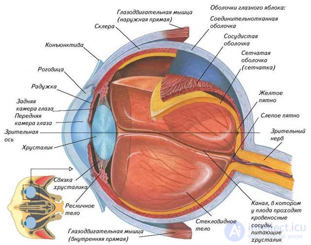
Eye structure
Visual perception begins with a projection of the image on the retina of the eye and the excitation of photoreceptors that transform light energy into nervous excitement. The complexity of the visual signals from the outside world, the need for their active perception led to the formation in the evolution of a complex optical device. This peripheral organ of vision is the eye.
The shape of the eye is spherical. In adults, its diameter is about 24 mm, in newborns - about 16 mm. The form the eyeball in newborns is more globular than in adults. As a result of this form of the eyeball, newborn children in 80-94% of cases have long-sighted refraction.
The growth of the eyeball continues after birth. It grows most intensely during the first five years of life, less intensely until 9–12 years old.
The eyeball consists of three shells — outer, middle, and inner.
The outer shell of the eye is the sclera or the protein shell. This is a dense opaque white fabric with a thickness of about 1 mm. In front of it, it is in front of the transparent cornea. The sclera in children is thinner and has a high tensile properties and elasticity.
The cornea in newborns is thicker and bulging By 5 years, the corneal thickness decreases, and its radius of curvature almost does not change with age. With age, the cornea becomes more dense and its refractive power decreases. Under the sclera is the choroid. Its thickness is 0.2-0.4 mm. It contains a large number of blood vessels. In the anterior part of the eyeball, the choroid enters the ciliary (ciliary) body and the iris (iris).
In the ciliary body is a muscle associated with the lens and regulating its curvature.
The crystalline lens is a transparent elastic formation shaped like a biconvex lens. The lens is covered with a transparent bag, along its entire edge, thin but very elastic fibers stretch towards the ciliary body. They are tightly stretched and hold the lens in a stretched state. The lens in newborns and preschool-age children has a more convex shape, is transparent and has greater elasticity.
In the center of the iris there is a hole-pupil. The size of the pupil changes, causing more or less light to enter the eye. The lumen of the pupil is regulated by a muscle located in the iris. The pupil of newborns is narrow. At the age of 6-8 years, the pupils are wide due to the predominance of the tone of the sympathetic nerves innervating the muscles of the iris. In 8-10 years, the pupil again becomes narrow and very vividly reacts to light. By the age of 12-13, the speed and intensity of the pupillary reaction to light are the same as in an adult.
The iris tissue contains a special dye-substance- melanin. Depending on the amount of this pigment, the color of the iris varies from gray and blue to brown almost black. The color of the iris is determined by the color of the eyes. In the absence of pigment (people with such eyes are called albinos), light rays penetrate into the eye not only through the pupil, but also through the tissues of the iris. Albino eyes have a reddish tint. They have a lack of pigment in the iris is often combined with lack of pigmentation of the skin and hair. Vision in such people is reduced.
Between the cornea and the iris, as well as between the iris and the lens, there are small spaces called the anterior and posterior chambers of the eye, respectively. They contain a clear liquid. It supplies nutrients to the cornea and lens, which are devoid of blood vessels. The eye cavity behind the lens is filled with a transparent jelly-like mass of the vitreous body.
The inner surface of the eye is lined with a thin (0.2-0.3 mm) very complex in structure, retina or retina sheath. It contains light-sensitive cells, called for their shape cones and chopsticks. The nerve fibers that extend from these cells come together and form the optic nerve, which is sent to the brain.
Functionally, they are apparatom muscles, protective devices, tears, eyelids and eyelashes.
The motor apparatus is formed by 6 muscles, of which 4 are straight and 2 are oblique. Rectus muscles rotate the eyeball around the transverse and vertical axis of the eye, oblique, around the axis passing in the anteroposterior direction. Consequently, the rectus muscles turn the eyeball left and right, up and down, and the oblique - down and in, up and in. All movements of the normal eyes of schoolchildren are only friendly.
The lacrimal apparatus consists of the lacrimal gland of a lobular structure, the lacrimal ducts and the lacrimal sac, or lacrimal lake. Lacrimal fluid washing the eyeball and the inner surface of the eyelids, protects them from drying out, protects against bacteria, due to its bactericidal properties. Eyelids and eyelashes are a kind of “sliding screens” that protect the eye from damage and dust, etc.
Hyperopia and myopia.
If the light rays passing through the pupil are refracted by the lens and focus on the retina, this is emmetropic vision (normal). If the rays of light, passing through the pupil, are refracted by the lens and focus before reaching the retina, this is myopia (myopia).
If the rays of light passing through the pupil are refracted by the lens and are focused behind the retina, this is hypermetrical vision (farsightedness).
Correction of myopic vision is carried out with negative lenses, with hypermetropic vision using positive lenses.
In addition to light, a person perceives color (red, yellow, green — primary colors). Loss of color perception - Daltonism.
Astigmatism
According to oculists, astigmatism is present in almost all the inhabitants of the Earth, but most of them (85%) have a barely noticeable one that does not affect visual acuity. The remaining fifteen percent have to suffer, try to correct this defect with special glasses (or lenses), or agree to an operation. What is this?
The word "astigmatism" consists of the Greek "stigme", which means the point, and the particle-negation "a". Thus, astigmatism is an eye disease in which there are no points at all. When astigmatism after refraction in the optical system of the eye, the light rays do not converge at one point, but are projected onto the retina in the form of several points, segments of different lengths, circles or ovals. As a result, instead of the normal image, something deformed and fuzzy is obtained. Moreover, a person suffering from astigmatism, equally bad sees both close and distant objects.
The reasons
The main cause of astigmatism is the irregular shape of the lenses of the optical system of the eye. Most often, the problem lies in the uneven curvature of the cornea, at least in the lens.
Normally, the cornea has a spherical shape, that is, its refractive power in the vertical and horizontal planes are the same. In astigmatism, the refractive forces of the cornea in these planes are different, for example, the cornea refracts vertically more strongly than horizontally.
Astigmatism is farsighted, short-sighted, and even combined: farsighted on one axis, short-sighted on the other.
Both adults and children suffer from it. In most cases, astigmatism is inherited and is called congenital. Acquired astigmatism usually develops due to gross cicatricial changes in the cornea after injuries and eye surgeries.
Age features of the visual analyzer.
Age-specific features of the visual analyzer are:
The smaller the child, the
1. Less visual acuity and lower threshold of sensitivity to the stimulus;
3. The less pronounced is the ability to refraction, the ability to perceive the power of the stimulus (the child blinks);
4. The faster the fatigue of the visual analyzer and the voltage of the Zinn ligaments develop;
The optic nerve of the left eye enters the visual center in the right hemisphere, the right eye in the visual center located in the left hemisphere.
This is a set of norms, conditions and requirements, which should be carried out to create optimal conditions for the activity of the visual analyzer.
1. Compliance with the norms of natural and artificial light.
2. Proper selection of furniture, taking into account the height of the child (the distance from the eyes to the table is 30-35 cm).
3. Compliance with the standards and requirements for watching television.
4. The correct dosage of visual loads (font for each age, cannot be read lying down, in a moving vehicle, keep distances, observe letter continuity standards: for students 6-7 years old 5-7 minutes, 7-10 years 10 minutes, 11-12 years 15 min, 13–15 years 20 min, 16–18 years 25–30 min Continuous reading: 6–7 years 5–10 min, 8–10 years 15–20 min, 11–15 years 25–30 min, 16 -18 years 35-45 minutes, in the intervals should rest the eyes for about 10 minutes).
The auditory analyzer is the second most important analyzer in providing adaptive responses and human cognitive activity, its special role in humans is associated with articulate speech.
Auditory perception is the basis of articulate speech. A child who has lost hearing in early childhood loses his speech ability, although his entire articular apparatus remains unbroken.
The auditory analyzer senses auditory waves that differ in height, frequency, and inner ear. Sound waves enter the outer ear, consisting of the auricle and the ear canal, go into the middle ear, consisting of the eardrum and 3 auditory ossicles, malleus, anvil, stirrup, then enter the inner ear, which includes a labyrinth, which consists of three parts : in the center is the vestibule, in front of it is a snail, consisting of 2.5 turns, behind are the semicircular canals. В центре улитки расположены рецепторы слухового анализатора – звуковоспринимающий аппарат- спиральный, или кортиев орган, представляющий собой слуховые волосики, ударяясь о которые звуковая волна преобразуется в электрический импульс, передающийся в слуховой нерв, который поступает в слуховой центр.
Слуховой анализатор включает вестибулярный аппарат, обеспечивающий удержание тела в пространстве.
Чем меньше ребенок:
1. Тем меньше пороги слышимости, наименьшая величина порогов слышимости, т.е. наибольшая острота слуха свойственная подросткам и юношам (14-19 лет)
2.Тем ниже острота слуха.
3.Тем быстрее развивается утомление слухового анализатора.
Гигиена слухового анализатора- это комплекс норм, условий и требований, направленных на охрану слуха, создание оптимальных условий для деятельности слухового анализатора, способствующих нормальному его развитию и функционированию.
1. Для слуха детей вредны чрезмерно сильные звуки. Это может привести к стойкому снижению слуха и даже полной глухоте.
2.Профилактика “ школьного шума”.
3.Речь учителя должна быть живой, богатой разнообразными интонациями, слова должны произноситься четко.
4.Правильная дозировка слуховых нагрузок.
5.Гигиеной слуха диктуются размеры учебного помещения.

Comments
To leave a comment
Human physiology, hygiene and age physiology
Terms: Human physiology, hygiene and age physiology